
|
|
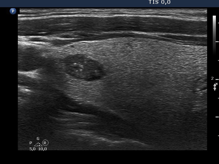
Intranodular hyperechogenic figures - case 975
|
|


Clinical presentation: A 40-year-old man was referred for evaluation of a nodular goiter detected on ultrasound screening.
Palpation: a firm nodule in the lower, isthmic part of the right lobe.
Functional state: euthyroidism with TSH 0.80 mIU/L.
Ultrasonography. The thyroid was echonormal. There were two nodules in the right lobe. The upper one was hypoechogenic and had punctate echogenic foci (microcalcifications) while the lower one was inhomogeneous, had cystic areas and presented back wall cystic figures.
Aspiration cytology of the upper lesion resulted in colloid goiter.