
|
|
Intranodular hyperechogenic figures - case conp 009
|
|


Clinical data: A 24-year-old woman requested an evaluation of thyroid dysfunction which has been detected by chance on routine laboratory investigation for two weeks. The GP administered 20 mg methimazole. She had no complaints suggesting hyperthyroidism.
Palpation: a moderately firm nodule in the left lobe.
Laboratory tests: TSH 8.72 mIU/L, FT4 8.51 pM/L, TSAb 0 U/L, anti-TPO 108 U/mL on daily 20 mg methimazole.
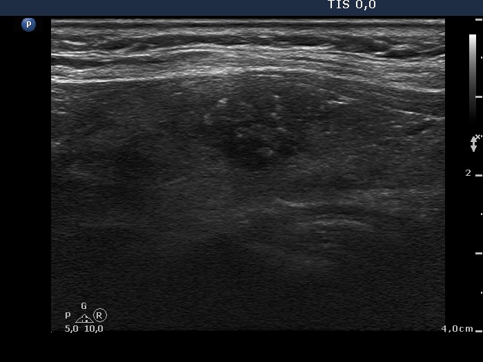
Ultrasonography. The thyroid was moderately hypoechogenic. There was a hypoechogenic nodule presenting hyperechogenic figures in the ventral part of the left lobe. The lesion had irregular, partly lobulated, partly blurred borders and presented intranodular blood flow.
Cytology resulted in Hürthle-cell tumor. Considering the ultrasound pattern, we raised the possibility of an oxyphilic variant of a papillary carcinoma.
Final diagnosis: Hashimoto's thyroiditis, Hashitoxicocis, thyroid nodule with the suspicion of malignancy. The methimazole therapy was discontinued.
Histopathology disclosed papillary carcinoma and Hashimoto's thyroiditis.
Comment. It is equivocal how to categorize the intranodular hyperechogenic figures in this case. They partly correspond to proliferation of connective tissue because as the video proves there were similarly bright lines and granules within the lesion. The issue is the coexistence of microcalcifications.