
Intranodular hyperechogenic figures - case conp 061 (ultrasonographic picture 6)
|
|

|
|
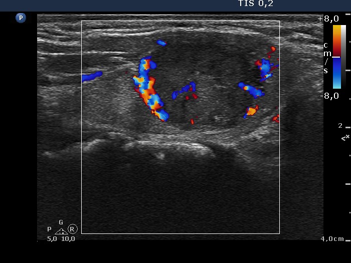
Right lobe, longitudinal scan, color Doppler mode. The lesion has perinodular and increased intranodular blood flow.