
Intranodular hyperechogenic figures - case cons100_012 (cytologic picture 1 of smear 2)
|
|

|

|
|
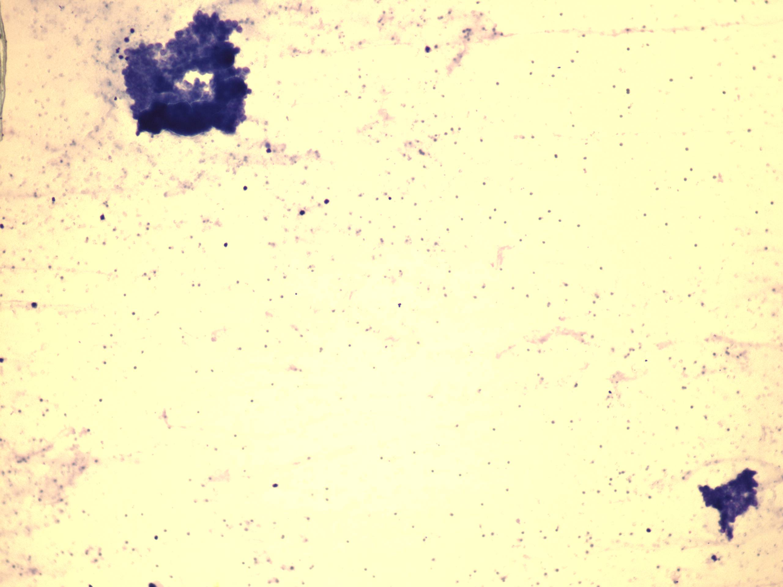
Wright-Giemsa staining, 100x. This smear was prepared from a small amount of brownish fluid. The image demonstrates the most cellular part of the smear.