Intranodular hyperechogenic figures - case cons100_012 (ultrasonographic picture 6)
|
|

|
|
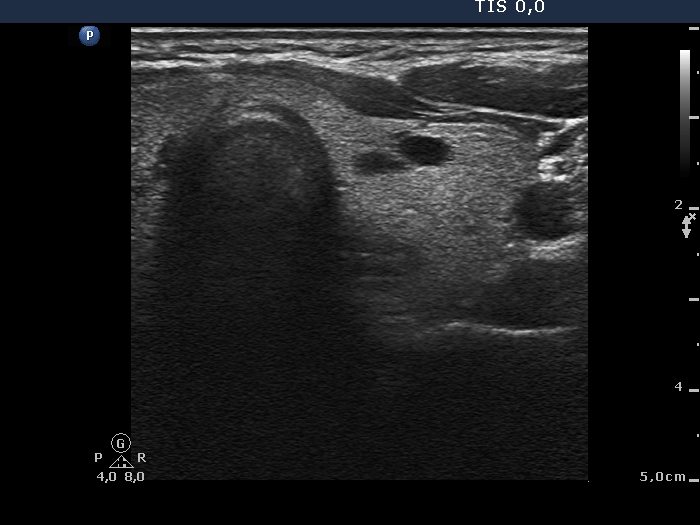
Left lobe lobe, transverse view. Two small cystic lesions are presented within an echonormal background.

Echogenic figures
|
|
|
|
Left lobe lobe, transverse view. Two small cystic lesions are presented within an echonormal background.