
|
|
The echogenicity of the nodule - case 2121
|
|


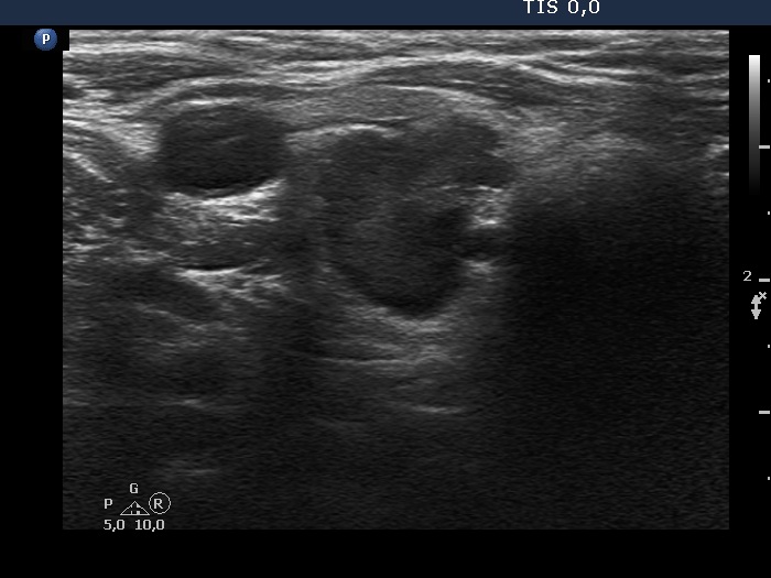


Clinical presentation: A 71-yr-old woman was referred for follow-up of an autonomously functioning adenoma, which has been known for 17 years. The patient had 'lump in the throat feeling'.
Palpation: a large, not firm nodule in the right lobe.
Laboratory test: TSH 1.65 mIU/L, FT4 14.1 pM/L.
Ultrasonography. The right lobe had a large isoechoic nodule. Dorsal and lower to this lesion there was a hypoechoic nodule, which presented irregular, lobulated margins. The hypoechoic nodule presented irregularly increased intranodular vascularization.
Cytology of the hypoechoic lesion resulted in benign, colloid goiter.
Comment.
- The hypoechoic nodule had irregular, lobulated margins.
- The categorization of the larger nodule depends on the definition of heterogeneity and the interpretation of the ventral hypoechoic part. If the latter is counted as a cystic area than the proportion of hypoechoic areas is below the 10% threshold and the nodule is not heterogeneous.