
|
|
The echogenicity of the nodule - case conp 049
|
|


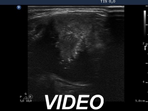


Clinical presentation: A 31-year-old woman was referred for evaluation of a nodular goiter which was known for a few months.
Palpation: a large, moderately firm nodule in the right lobe and in the isthmus.
Functional state: euthyroidism (TSH 0.98 mIU/L).
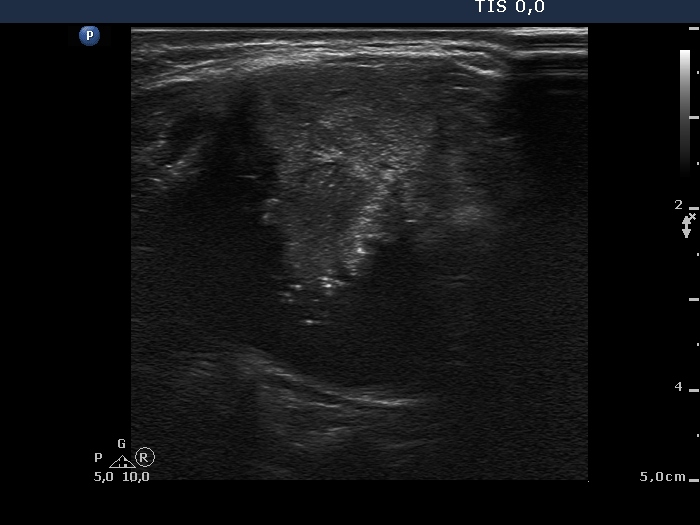
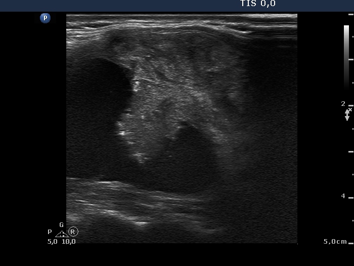
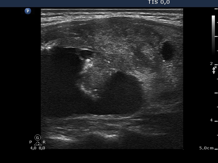
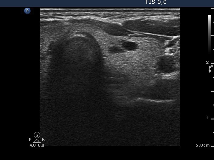
Ultrasonography: The thyroids was echonormal. A large, peripheral-type cystic nodule occupied almost the entire right lobe. The solid part was heterogeneous with equal portion of hypoechoic and echonormal parts and contained microcalcifications. There was no intranodular vascularization. The left lobe presented several small cystic and moderately hypoechogenic lesions.
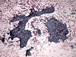
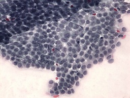
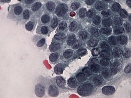
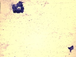
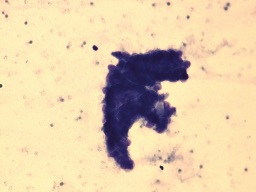
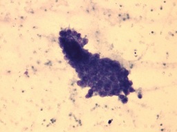
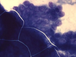
The nodule was aspirated three-times. The images of two smears are presented. Cytological diagnosis: papillary carcinoma.
Histopathology disclosed papillary carcinoma in the right lobe while benign hyperplastic nodules in the left thyroid.
Comments:
- As regards the ultrasound presentation there were two features which increased the risk of malignancy: the presence of microcalcifications and the peripheral-type of the cyst. On the other hand, two other features decreased the likelihood of carcinoma: the cyst presented blunt angle and the vascularization was not increased.
- Although the upper cytological images are clearly diagnostic, the lower images may be more edifying. In significant proportion of cystically degenerated papillary carcinomas, we gain only small fragments of cells, and it is very difficult to give a correct diagnosis.
- The cystic lesions presented comet-tail artifact while two moderately hypoechogenic small nodules did connective tissue.
.