
|
|
The echogenicity of the nodule - case conp 058
|
|


Clinical data. A 22-year-old woman was investigated for a year because of nodular goiter in another hospital. The cytology was of limited value because of small number of cells but no signs of malignancy was found. The patient was referred to us for follow-up investigation. She had no complaints.
Palpation: a firm nodule in the right lobe and an enlarged, not freely moveable lymph node above the right thyroid.
Functional state: euthyroidism with TSH-level 1.94 mIU/L.
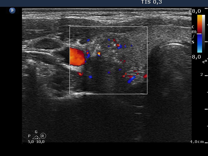
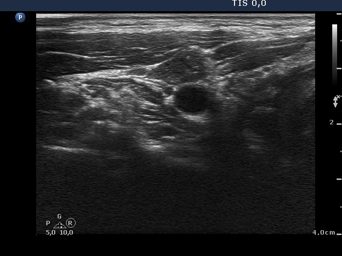
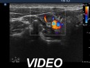
Ultrasonography. There were two nodules in the right lobe. The upper contained multiple punctate echogenic foci while the dorsal one had irregular, lobulated margins. Both lesions presented irregularly increased intranodular vascular pattern. The lymph node above the right thyroid presented irregular vascularization and punctate echogenic foci and lacked hilum. The ventral lesion was heterogeneous, containing nearly equal proportions of iso/hyperechoic and hypoechoic parts. The dorsal lesion was deeply hypoechoic.
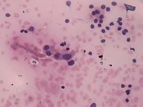
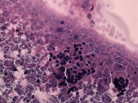
Cytology was performed from the nodule and the lymph node. Only one of the five smears was diagnostic. This smear contained two cell populations. The predominant type corresponded to a benign colloid goiter. Small number of atypical cells were also found. They were enlarged, have abundant cytoplasm and prominent nucleoli. There were only heterogeneous lymphoid cells in the smear of the lymph node.
Combined sonographic-cytological diagnosis: suspicion of malignancy. Papillary carcinoma should be considered first.
Histopathology: multifocal papillary carcinoma in the right lobe with metastasis to the neck lymph nodes.
Comments.
-
There are two cell populations on the smear which is a rare finding and emphasizes the role of thorough analysis of the smears.
-
The cytological pattern itself was not enough to raise the possibility of a carcinoma. It corresponded to AUS of Bethesda system. Nevertheless, taking the sonographic pattern into account the preoperative diagnostic could raise the possibility of carcinoma.