
|
|
Extrathyroidal spread - case 2083
|
|


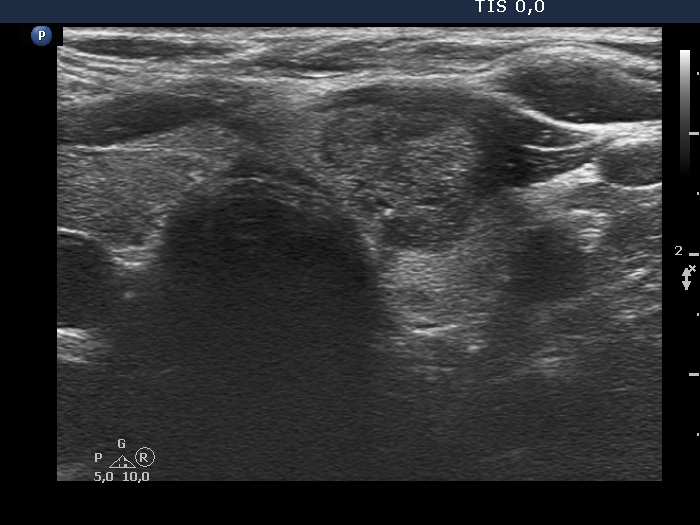


Clinical presentation: A 50-yr-old woman was referred for aspiration cytology of a nodular goiter which has been discovered on evaluation of elevated blood pressure.
Palpation: no abnormality.
Laboratory test: TSH 0.71 mIU/L.
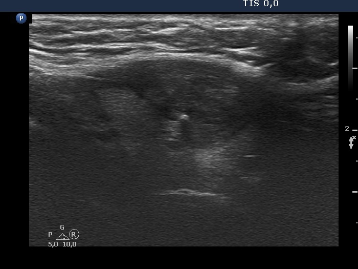
Ultrasonography. The thyroid was echonormal. There was a heterogeneous, hypoechoic-isoechoic mass in the ventral part of the left lobe. The mass caused protrusion in the ventral surface of the lobe. The dorsal borders of the mass were undulated.
Cytology was performed from the lesion in the left lobe and resulted in benign, colloid goiter.
Comments.
-
Occasionally, it is difficult or even impossible to judge whether a lobulated surface is caused only by the presence of multiple discrete lesions or certain parts of a lesion are in fact pathologically lobular. The clue is the thorough analysis of video. In this case pathological lobulation is not likely.
-
The nodule presents all three signs which can raise the suspicion of extrathyroidal extension: the contour is abutting and bulging and the capsule of the thyroid is discontinuous.