
|
|
Extrathyroidal spread - case 2128
|
|


Clinical presentation: A 48-year-old woman was referred for aspiration cytology. The patient has been known harboring a nodule for two years.
Palpation: a moderately firm nodule in the right lobe.
Laboratory test: TSH 2.01 mIU/L.
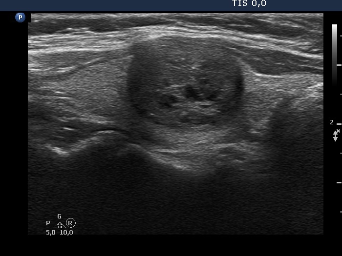
Ultrasonography. The thyroid was echonormal. There was a moderately and a minimally hypoechoic nodule in the right lobe, in the middle and lower part, respectively. The former had intranodular echogenic figures, most of them belonged to back wall cystic figures except for a few more bright ones which could be microcalcifications.
Cytology was performed from both nodules and resulted in benign cystic-colloid goiter and in colloid goiter, middle and lower nodule, respectively.
Comment. The lesion in the middle part presented all three possible signs on which extrathyroidal extension can be suspected, i.e. the contours were abutting, and bulging and the capsule of the lobe was not continuous. The lower nodule displayed abutting and the capsule of the lobe was also discontinuous in the event of this lesion.