
Extrathyroidal spread - case 2138 (ultrasonographic picture 8)
|
|

|
|
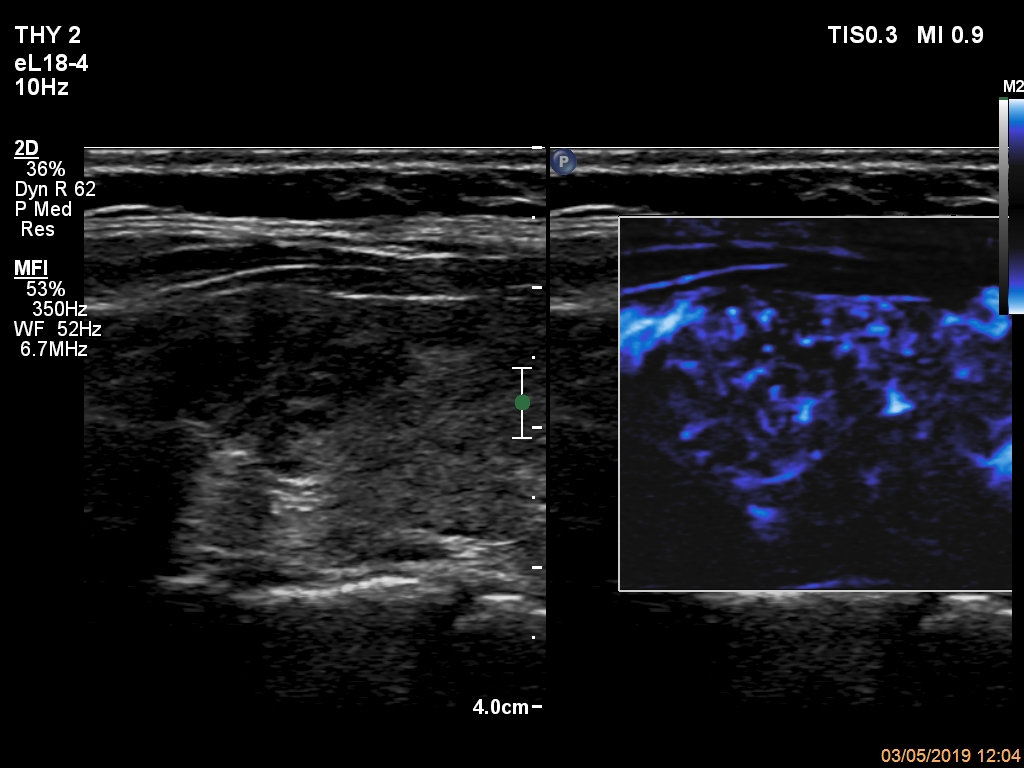
Left lobe, longitudinal scan, microflow imaging. The vascularization of the upper discrete hypoechoic area (left in the image) does not differ from that of the other parts of the lobe.