
|
|
Extrathyroidal spread - case conp 026
|
|


Clinical data: A 66-year-old woman was referred for evaluation of an elevated parathormone level.
Palpation: Both lobes were nodular.
Functional state: euthyroidism (TSH 2.50 mIU/L). Parathormone 162.2 pg/mL, calcium 2.07 mM/L, phosphate 1.1 mM/L.
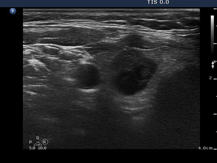
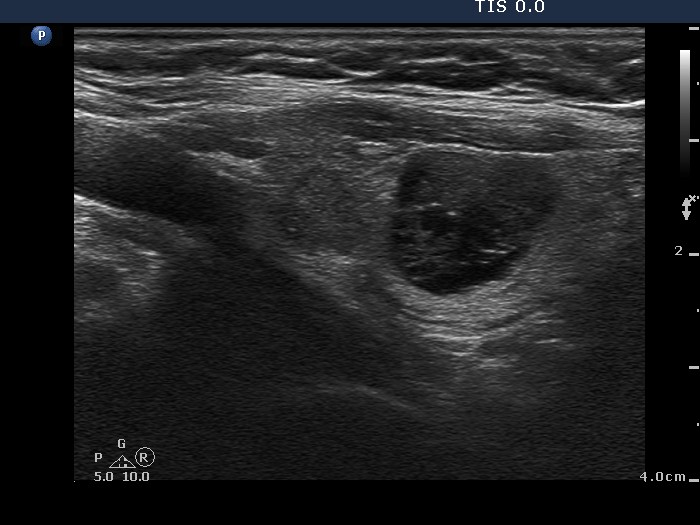
Ultrasonography. The thyroid was echonormal. There were multiple nodules of different echogenicities in the right lobe. There was hypoechogenic nodule that presented irregular borders and hyperechogenic granules in the left lobe.
Cytology was performed the lesion in the left lobe and resulted in suspicion of carcinoma. Wash-out thyroglobulin resulted in 155 ug/L, serum thyroglobulin did in 21.6 ug/L. Serum calcitonin resulted in 1.43 mM/L (normal value 0-3.36). Our final diagnosis was suspicion of papillary carcinoma.
Histopathology disclosed papillary carcinoma in the left lobe while hyperplastic nodules in the right lobe. The tumor proved to be a T4a tumor.
Comment. The malignant nodule presented all three possible signs which might suggest extrathyroidal extension: abutment, bulging and discontinuation of the thyroid capsule. This is one the few examples in which we can clearly diagnose extrathyroidal extension on ultrasound examination.