
|
|
Extrathyroidal spread - case conp 051
|
|


Clinical presentation: A 69-year-old woman came to a regular follow-up investigation. We examined her 5 years ago when she had a nodule with the dimensions of 8x8x13 mm. FNA was performed and it gave a benign result. The patient had no complaints.
Palpation: a moderately firm nodule in the right lobe.
Results of blood tests: euthyroidism (TSH-level 1.05 mIU/L).
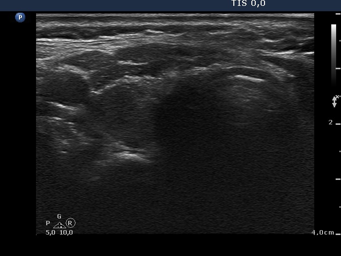
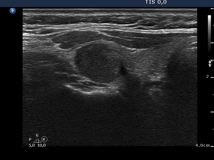
Ultrasonography: The thyroid was echonormal. A moderately hyperechogenic, inhomogeneous nodule was present in the right lobe with the dimensions of 10x11x13 mm. It means that the volume of the nodule increased from 0.4 ml to 0.75 ml in 5 years. The nodule presented increased intranodular blood flow.
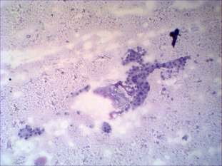
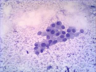
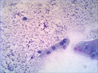
Cytological picture. There was no colloid in the background. A few atypical clusters were present. A great proportion of follicular cells exhibited nuclear groove, a few cells did inclusion.
Clinical-cytological diagnosis: papillary carcinoma.
Histopathology: multifocal papillary carcinoma.
Comments.
-
We reviewed the original smears which have gained for 5 years. There were only a few cells on the smear without any atypia. The correct diagnosis would be not diagnostic.
-
The borders of the nodule these are blurred.
-
The dorsal contour of the nodule is in part in contact with the surface of the thyroid. The issue is the degree of abutment. Video proves that the degree of abutment exceeds 25% in certain sections.