
|
|
Halo sign and vascular pattern of nodules - case 1744
|
|

Clinical data: A 71-year-old woman was referred for evaluation of a painless mass in the left thyroid which appeared 3 weeks ago.
Palpation: a firm nodule in the left lobe.
Laboratory test: TSH 0.91 mIU/L.
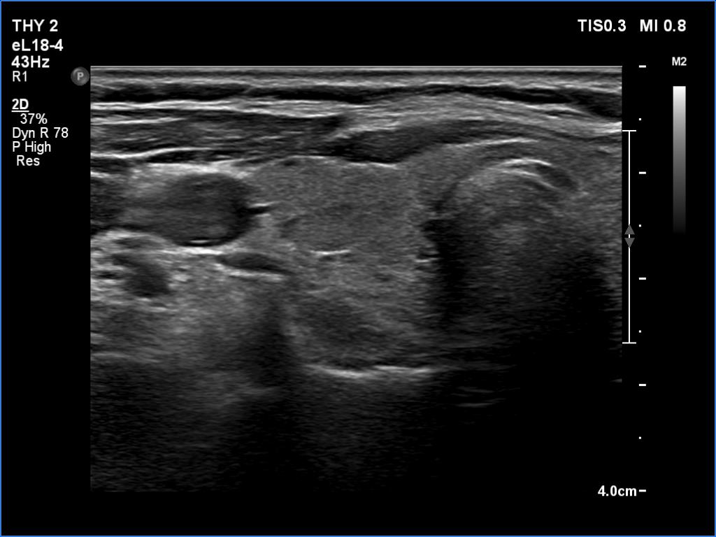
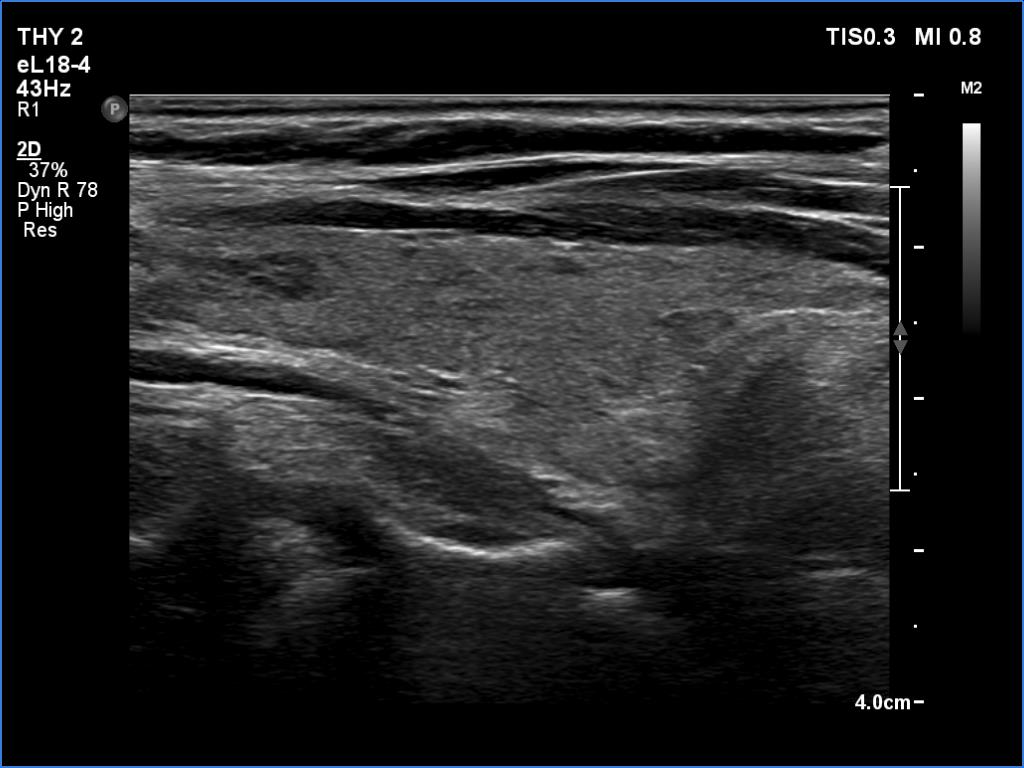
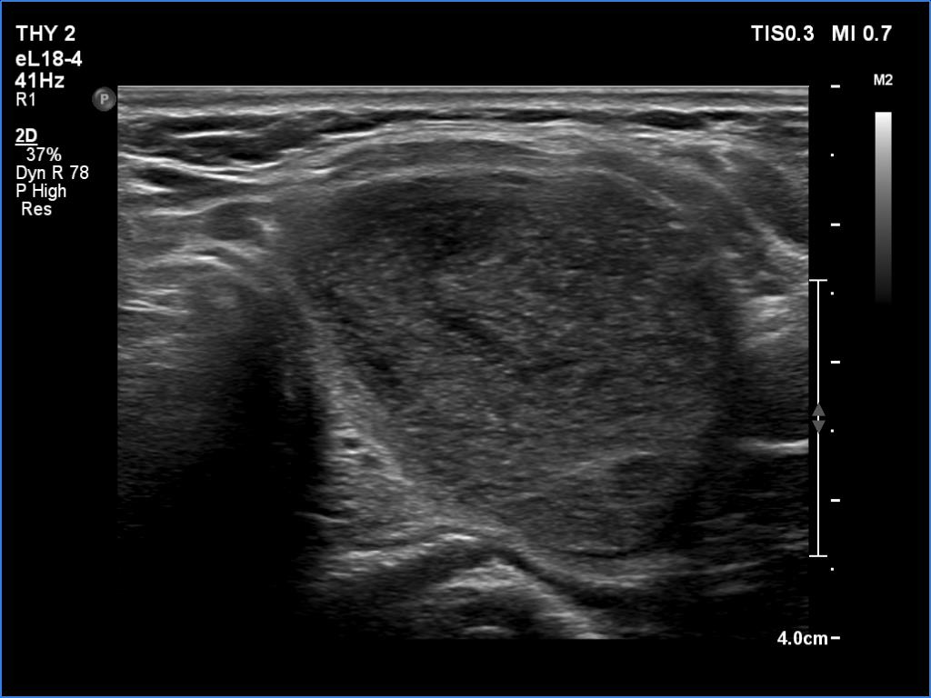
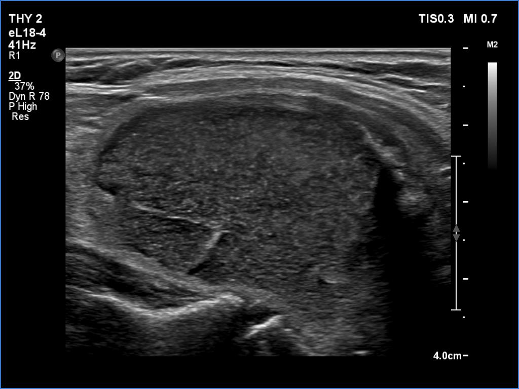
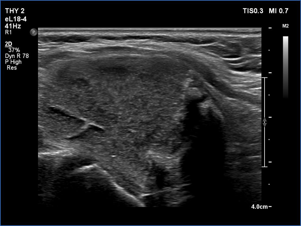
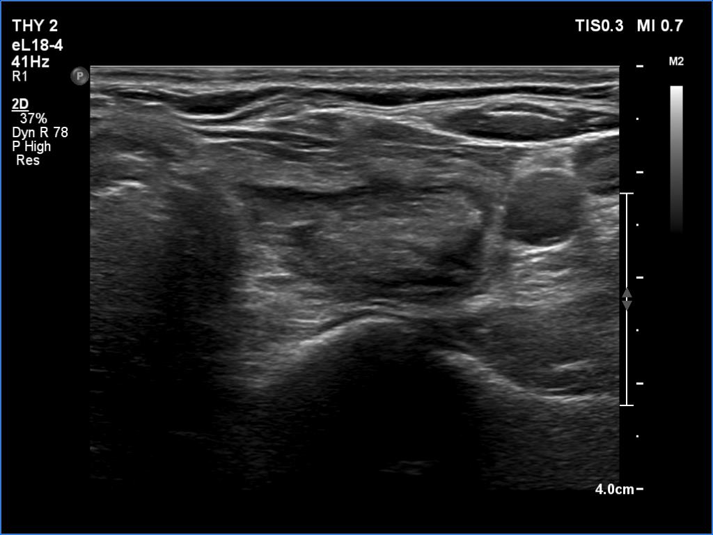
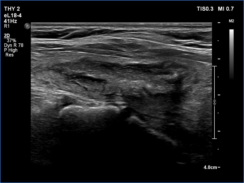
Ultrasonography. The thyroid was echonormal. There were several hypoechoic areas in the right lobe. The left lobe had a large minimally hypoechoic nodule with several tiny cystic areas. The lesion had numerous back wall figures.
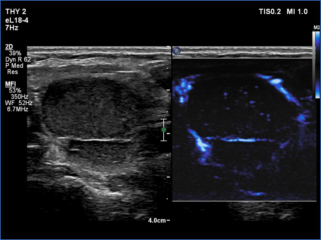
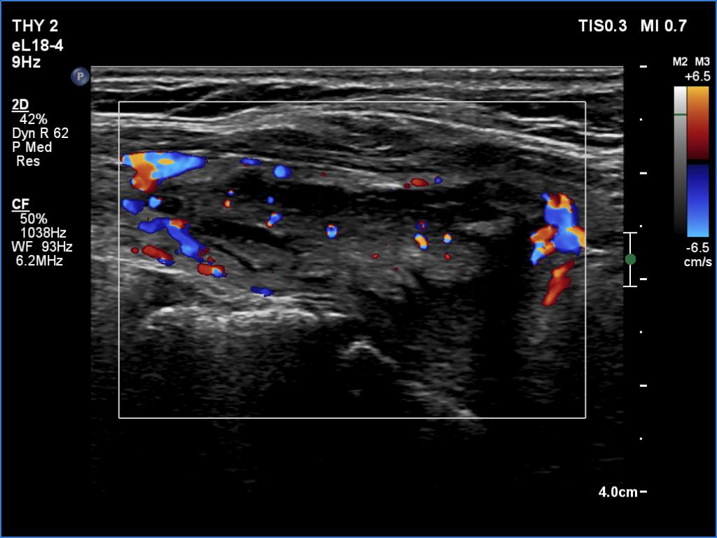
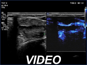
In the first part of the ultrasound examination, when the transducer was continuously moving over the thyroid gland, it was not detectable that the contents of the nodule were showing flow. When we stopped the transducer, it became clear that the seemingly solid mass was actually a dense liquid in a continuous flow. On Doppler examination, 'circulation' was visible in continuously changing places.
If it had really been about blood vessels, according to the cycle of circulation,
it would have disappeared once, at other times the flow would have reappeared, but at the same time we would have always seen it in the same place.
14 mL thick yellow fluid was aspirated. Cytology resulted in cystic fluid only, Bethesda I category.
Comment.
This case illustrates two rare phenomena. On the one hand, a dense liquid may appear to be a solid tissue. On the other hand, the Doppler assay actually detects fluid flow. In the vast majority of cases, of course, this displays a circulation in the blood vessels, but in the case of cysts, when pressure is applied to the fluid with the probe, a flow is created. In the case of flow in blood vessels, circulation is always seen in the same place, while in the case of flowing cystic fluid, the flow can be detected in constantly changing places.