
Halo sign and vascular pattern of nodules - case 2254 (ultrasonographic picture 4)
|
|

|
|
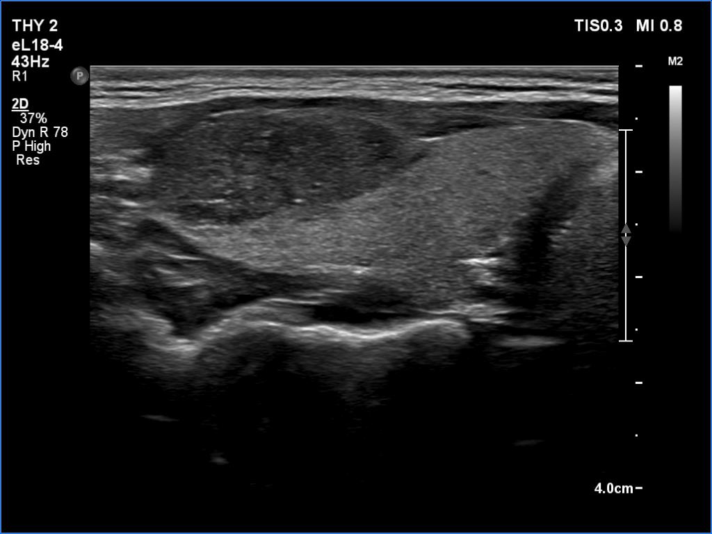
Right lobe, another longitudinal view. In this section, punctate echogenic foci can be revealed. However, the brighter granules are related to ventral cystic areas which means that these are back wall figures.