
Halo sign and vascular pattern of nodules - case 444 (ultrasonographic picture 2)
|
|

|
|
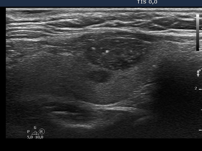
Right lobe, longitudinal scan. The nodule does not display halo sign. There are two larger granules in the lesion. The brighter in the central part could be a microcalcification but as the video proves, similarly bright hyperechogenic lines are also present. Moreover, the granules are found dorsal to tiny cystic areas. Therefore, the large and bright granule is very likely caused by back wall posterior enhancement.