
Halo sign and vascular pattern of nodules - case 738 (ultrasonographic picture 1)

|
|
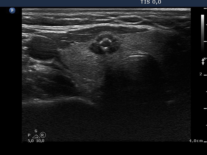
Right lobe, transverse scan. There is a hypoechoic nodule which has irregular borders. There are punctate echogenic figures within, these correspond to microcalcifications. Although the lesion bulges, this has minimal relevance in the absence of abutting contours.