
The shape of the nodule - case 2052 (ultrasonographic picture 2)
|
|

|
|
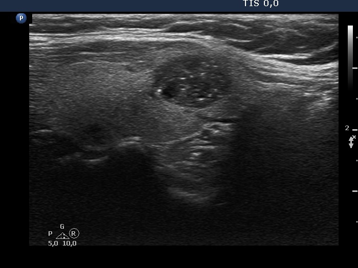
Left lobe, longitudinal scan. Considering this view, the interpretation of the echogenic figures has to changed. Most figures are related to ventral cystic areas which means that great proportion of these echogenic figures are caused by back wall posterior enhancement.