
|
|
The shape of the nodule - case 2122
|
|


Clinical presentation: A 74-yr-old woman was referred for evaluation of a multinodular goiter which was detected by chance on carotid Doppler examination.
Palpation: no abnormality.
Laboratory tests: TSH 1.67 mIU/L.
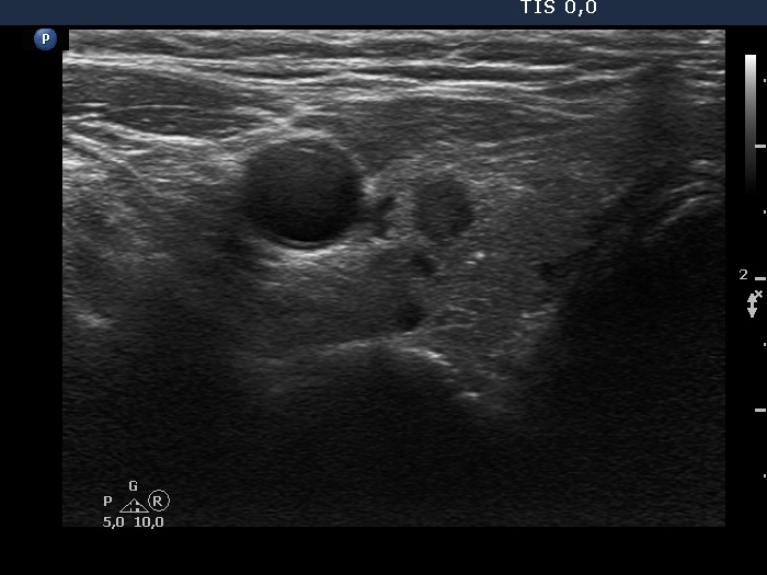
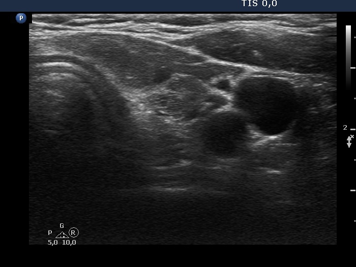
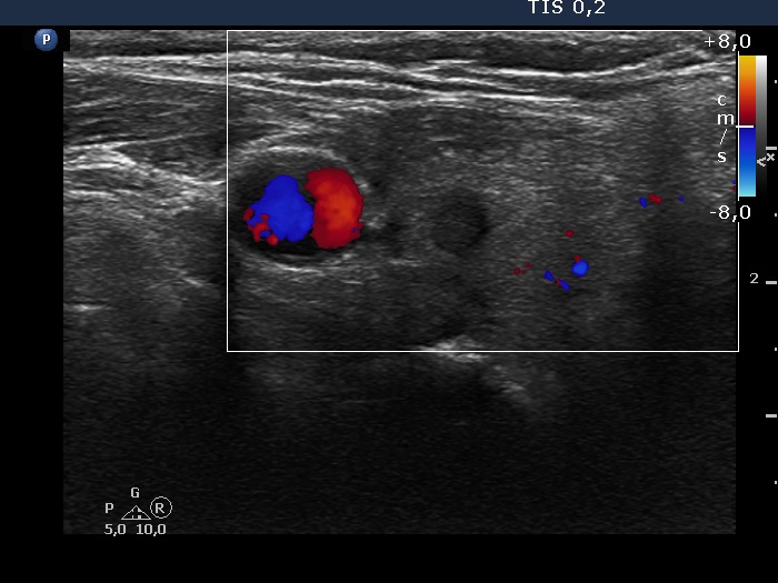
Ultrasonography. The thyroid was minimally hypoechoic and has several discrete lesions. A hypoechoic one in the left lobe showed both taller-than-wide and taller-than-long shape. There was a dominantly solid nodule in the lower part of the right lobe. There was a hypoechoic nodule in the upper, ventromedial part of the left lobe. This was the only lesion which was larger than 1 cm. This had a bit elongated hyperechoic figure.
Cytology of the nodule in the left lobe resulted in benign, follicular proliferation.
We performed aTPO determination which resulted in normal value (1.5 U/mL).
Suggestion: ultrasound in three years.
Comment. The pattern was suspicious of Hashimoto's thyroiditis. Nevertheless, in elderly persons the echogenicity normally decreases with age and the presence of small, discrete lesions is again a common finding.