
|
|
TIRADS - case 1649
|
|


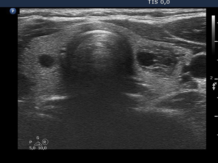


First examination (first and second rows of images):
Clinical presentation: A 27-year-old woman requested evaluation of a neck lump which was discovered by herself. She has felt neck discomfort for several weeks.
Palpation: an elastic nodule in the left lobe.
Result of blood test: TSH 1.32 mIU/L, FT4 16.0 pM/L, aTPO 13 U/mL.
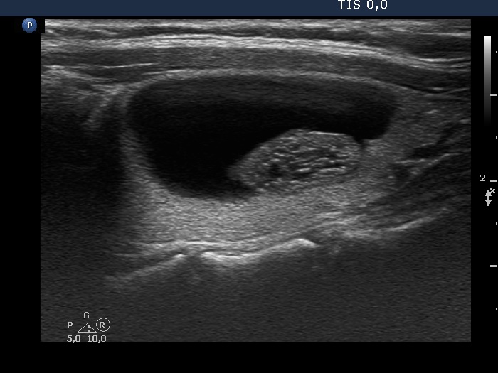
Ultrasonography. The thyroid was echonormal. There was a smally cystic area in the right lobe, while a peripheral-type cystic nodule in the left lobe. The latter presented numerous echogenic figures which were caused by back wall posterior enhancement. The dimensions of the nodule were 21x15x33, width, depth, length, respectively.After aspirating 5 mL serous fluid, aspiration cytology was performed from the solid part which resulted in benign cystic lesion.
First session of sclerotherapy (third row of images):
Clinical presentation. The complaints of the patient ceased and did not recur in the upcoming months. However, four months after the previous examination she noticed that her cyst has refilled.
Palpation: an elastic nodule in the left lobe.
Result of blood test: TSH 5.61 mIU/L.
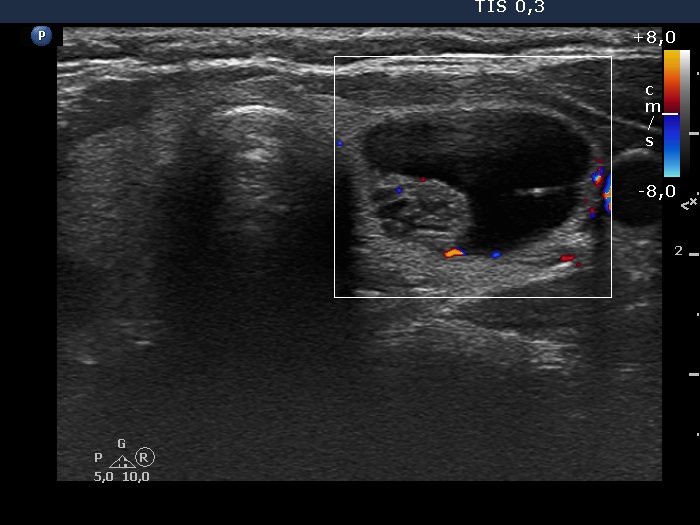
Ultrasonography. The pattern was the same as what we saw at the first examination. The dimensions of the nodule were 19x15x33, width, depth, length, respectively. The volume of the lesion was 5.44 mL.Two mL brown fluid was aspirated, then 0.8 mL ethanol was injected.
Third session of sclerotherapy (fourth row of images):
Clinical presentation. The left side of the neck became sore for four days. At the second session 2.5 mL brown fluid was removed and 1.4 mL ethanol was administered.
Palpation: The left lobe was a bit sensitive on palpation.
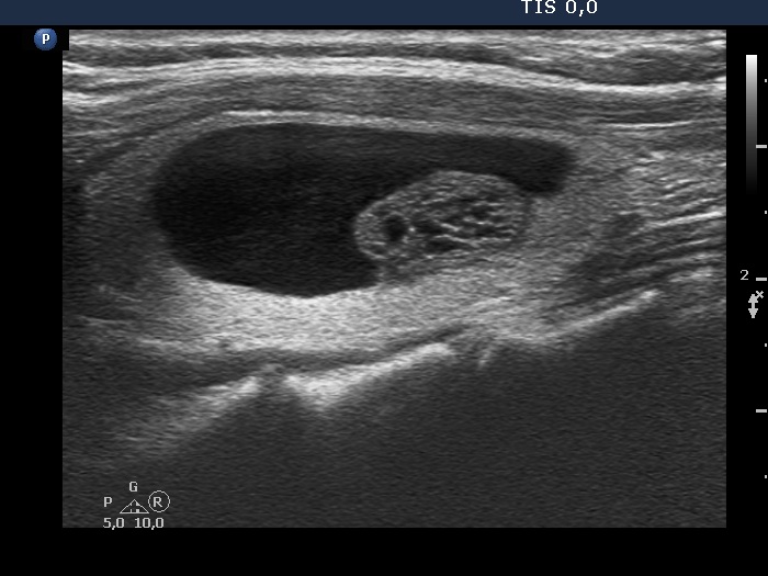
Ultrasonography. The dimensions of the nodule were 10x7x14 mm, width, depth, length, respectively.
This time 0.5 mL ethanol was injected.
Three months after the sclerotherapy (fifth row of images):
Clinical presentation. The patient came to routine follow-up visit. She has lost her husband for three months and thereafter diffuse complaints including weight loss, anxiety and sleep disturbance had evolved.
Palpation: unchanged.
Result of blood test: TSH 5.61 mIU/L.
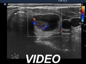
Ultrasonography. The right cystic nodule has in part refilled. Otherwise, the presentation remained unchanged. The dimensions of the nodule were 10x7x14 mm, width, depth, length, respectively. The volume of the lesion was 0.51 mL, i.e. one-tenth of the original.Suggestion: ultrasound in 6 months.
Comment. The pattern of the solid part at the first examination and at the first session is remarkable, it is very close to a spongiform pattern.