
|
|
TIRADS - case 60
|
|


Clinical presentation: A 64-year-old woman was referred for a follow-up examination. Aspiration cytology was performed from a lesion with a maximal diameter of 8 mm ten years ago and resulted in benign lesion. The patient had no complaints.
Palpation: a small, not firm nodule in the right lobe.
Functional state: euthyroidism with TSH 0.73 mIU/L.
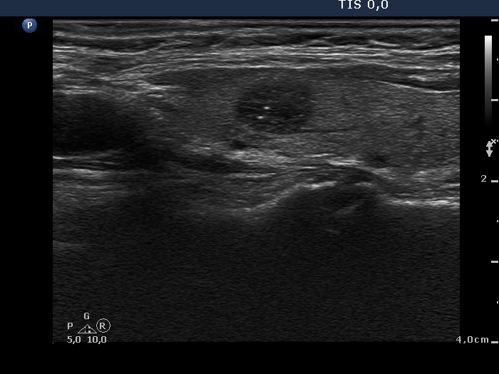
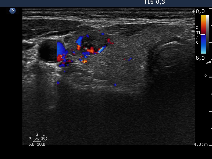
Ultrasonography revealed a deeply hypoechogenic nodule which had punctate echogenic foci. At the time of the examination, we interpreted these figures as microcalcifications. The nodule did not present a halo sign while did a combined perinodular and intranodular vascular pattern.
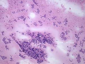
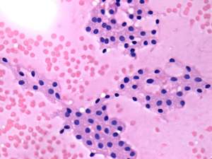
Cytological report: suspicion of a Hürthle-cell tumor.
Histopathology disclosed Hürthle-cell adenoma.
Comments.
-
The cytological pattern itself might correspond to either a hyperplastic nodule or an oxyphilic adenoma.
-
It is difficult to decide how to classify the intranodular echogenic granules. These are partly related to cystic areas, moreover echogenic lines are also present. The latter are less bright than the granules. I think we would overestimate our options if we made a clear commitment in this case. In any case, re-evaluating the case, I would be more cautious: in the case of intranodular echogenic granules, in addition to the back wall figures, microcalcifications should also be considered.
- The nodule should be classified as an EU-TIRADS 5 lesion due to the deeply hypoechoic pattern.