
Subacute granulomatous thyroiditis - case 1514
Examination a year after the first visit (ultrasonographic picture 8)
|
|

|
|
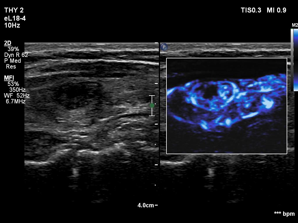
Left lobe, longitudinal scan, microflow imaging. The lobe and the discrete lesion are rich in vessels.