
Graves' disease - case 530
12 months after initial investigation (ultrasonographic picture 4)
|
|

|
|
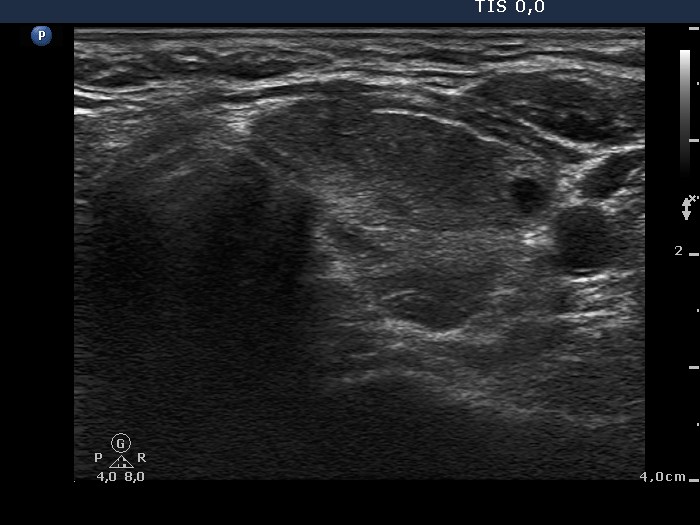
Left lobe, transverse scan. The left lobe is composed of discrete hypoechogenic areas and a central echonormal part. The pattern is identical to that seen at the first examination.