
|
|
Graves' disease - case conp 033
|
|


Clinical presentation: A 27-year-old man requested a second opinion. He has been treated for hyperthyroidism for 6 months.
Palpation: Both lobes were enlarged and moderately firm. There was no palpable nodule.
Results of blood tests: hypothyroidism on daily 20 mg propylthiouracil (TSH 8.81 mIU/L, FT4 8.01 pM/L).
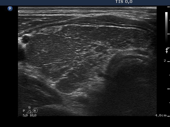
Ultrasonography. A diffusely hypoechogenic thyroid was found with extensive fibrosis. There was an echonormal nodule in the upper dorsal part of the left lobe. The nodule showed taller-than-wide shape and halo sign while the presence of perinodular blood flow was equivocal.
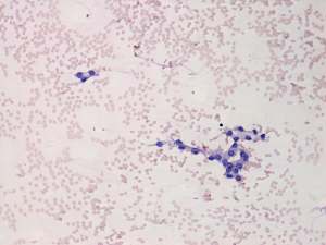
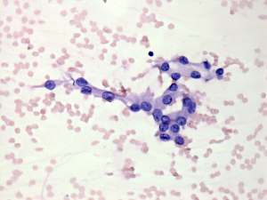
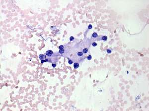
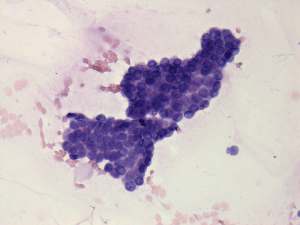
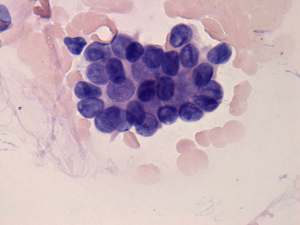
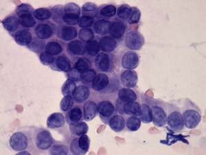
Cytology was performed from the hyperechogenic nodule and resulted in suspicion of papillary carcinoma. (The two rows of cytological images represent two different smears.)
Histopathology: diffuse goiter corresponding to Graves' disease and an encapsulated papillary carcinoma corresponding to the nodule. The tumor broke not only through the capsule but spread extrathyroidal.
Comments:
-
The extensive fibrotic change is a not rare situation in the case of Graves' disease. We can demonstrate the possible misinterpretation of granular presentation of fibrosis - see comment to ultrasonographic picture.
-
Malignancy occurs only exceptionally in an echonormal nodule except for autoimmune thyroid disorders. We must be aware that the echo structure of a nodule may be influenced by the echo structure of the non-nodular thyroid. Therefore, echonormal nodules are targets of cytological investigation in hypoechogenic thyroids, i.e. Graves' disease and in Hashimoto's thyroiditis.
-
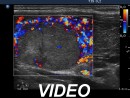
The presence of perinodular blood flow is doubtful because the nodule is avascular while the vascularization of the extranodular part is extremely increased. Nevertheless, the nodule proved to be encapsulated on histopathology.
-
The cytological pattern of smear-1 corresponds to atypia of unknown significance.
-
Although the pattern in smear-2 is almost pathognomonic and diagnostic, we do not give a definite diagnosis of papillary carcinoma if the nuclei lack inclusions.
- The histogram value of the nodule proved to be 74.8, a value which is less than the average of the normal parenchyma, therefore in absolute sense, this is a minimally hypoechoic nodule. In relative sense, compared to the non-nodular part of the particular case, the nodule is hyperechoic.