
Lymphocytic thyroiditis - case 325 (ultrasonographic picture 6)

|

|

|
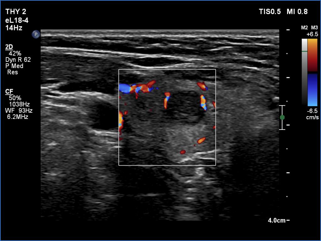
Middle part of the right lobe, transverse scan, color Doppler mode. There are two vessels running at the border of the nodule.


