
Lymph nodes in the neck - Case 123 (ultrasonographic picture 4)
|
|

|
|
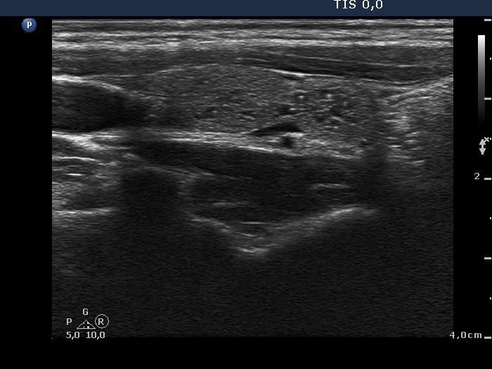
Right lobe, another longitudinal scan. At first sight, the lesion in the lower part contains numerous microcalcifications. However, similarly bright hyperechogenic lines can be found, too. Moreover, several granules have a short dorsal tail. So, the differential diagnostic involves beside microcalcification, connective tissue and colloid crystals, as well.