
|
|
Lymph nodes - case 1675
|
|


Clinical data: A 36-year-old woman requested evaluation of a neck mass in the right submandibular area. She has been aware of this mass since early childhood. A few weeks after an upper respiratory infection, she noticed that the neck mass had enlarged and then spontaneously retreated a week later.
Palpation: There was a firm mass in the right submandibular area. The right thyroid lobe had firm nodule.
Laboratory tests: TSH 0.99 mIU/L, FT4 17.3 pM/L, aTPO 167 U/mL.
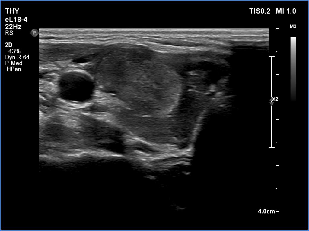
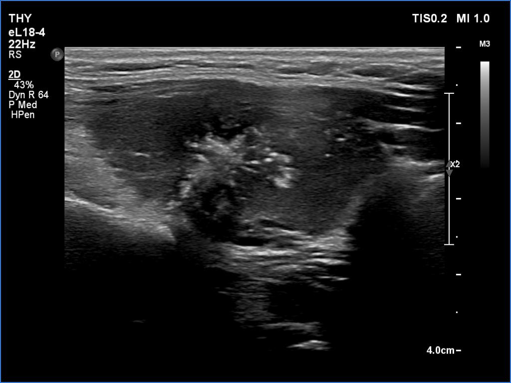
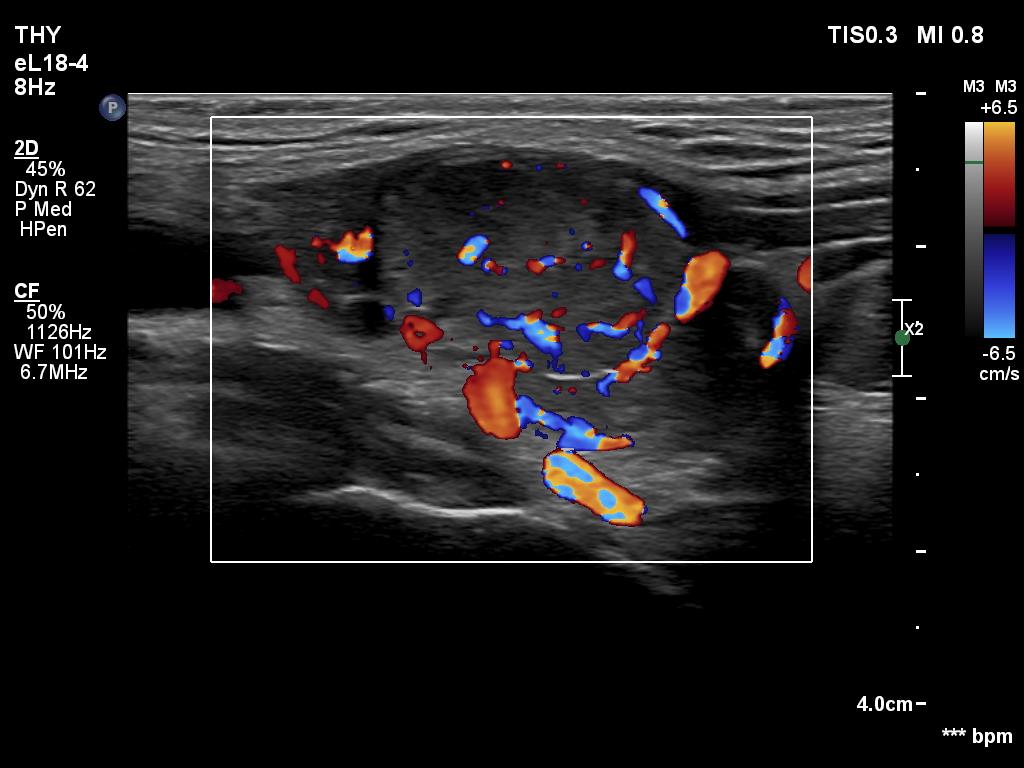
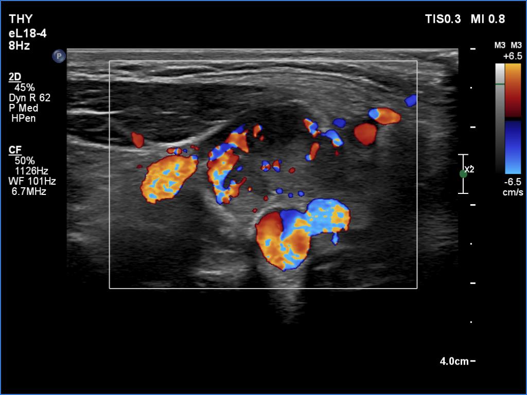
Ultrasonography. The thyroid was hypoechoic. The right lobe had a heterogeneous, dominantly minimally/moderately hypoechoic nodule in the ventrolateral part which had irregular borders and both perinodular and intranodular vascularity. There was another, hyperechoic star-like lesion in the central part of the right lobe. This lesion presented microcalcifications and was avascular. The left lobe was homogeneously hypoechoic.
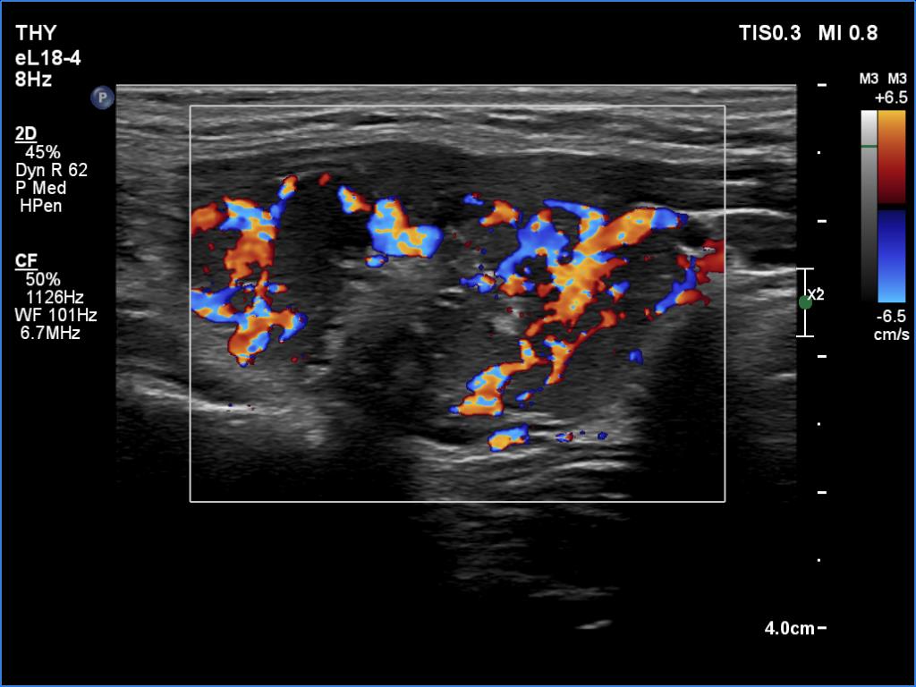
According to the palpable mass in the right submandibular area, there was a heterogeneous lesion which has hypoechoic and echonormal parts. The mass had irregular margins and showed vascularity. The distance between the submandibular mass and the thyroid was more than 35 mm.
Cytology resulted in Hashimoto's thyroiditis, and suspicion of oxyphilic variant of papillary cancer, the complex, dominantly minimally/moderately hypoechoic nodule in the ventrolateral part and the star-like hyperechogenic lesion. There were compact groups composed of cells with abundant cytoplasm and lymphoid elements in the smears gained from the submandibular mass. The cytological presentation corresponded to Hashimoto's thyroiditis. Wash-out thyroglobulin resulted in 172 ng/mL.
Total thyroidectomy was performed. Histopathology: disclosed papillary cancer according to the hyperechoic lesion in the right lobe. No other nodule was described in the thyroid. Hashimoto's thyroiditis was found in the extranodular thyroid and in the submandibular mass, as well.
Comments.
-
Although, the patient' history had little influence on the preoperative diagnosis in this case, it could have been of extreme importance. The presentation of the neck mass is highly suspicious of a metastatic lymph node, as was the cytological pattern: groups of epithelial cells within a lymphoid cell population - what else is required to consider a metastasis in a lymph node? (In addition to, the patient had a papillary cancer in her thyroid lobe.) But we had to take into account that this mass has been already present for decades.
-
It is worth noting the similarity of the pattern of the ectopic tissue and the thyroid.