
|
|
Parathyroid lesions - case 742
|
|


Clinical data: On routine laboratory blood test, an elevated serum calcium level, thereafter an elevated parathormone level (142 pg/mL) were detected in a 64-year-old woman 5 years ago. Further investigation of this was then interrupted for unknown reason. This time, the GP initiated repeat examination because the patient suffered from three bone fractures in the past one year.
Palpation: no abnormality.
Laboratory tests: TSH 0.98 mIU/L, parathormone 221 pg/ml, serum calcium 3.04 mM/L, phosphorus 0.75 mM/L.
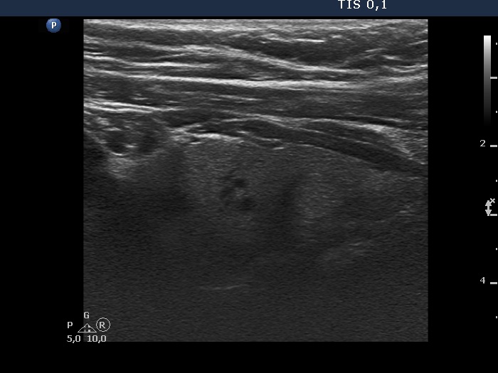
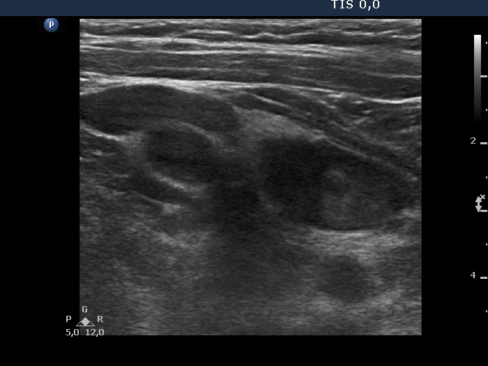
Ultrasonography. The thyroid was echonormal and had several insignificant hypoechoic lesions. There was a hypoechoic mass lower and dorsal to the lower pole of the left lobe.
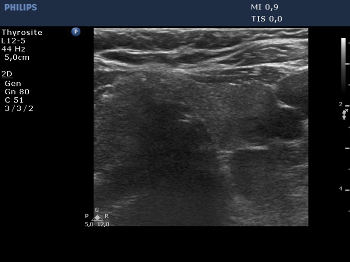
Aspiration cytology of the lesion was performed. It was doubtful whether we reached the parathyroid during aspiration. We gained three smears on which typical follicular cells were found in colloidal background.
Additional tests: Wash-out thyroglobulin resulted in 203 ng/mL, wash-out parathormone did in 1033 pg/mL.
MIBI scintigraphy disclosed an enlarged left lower parathyroid.
Histopathology: parathyroid adenoma.
Comment.
-
Targeting and hitting a lesion which is lower to the lower pole of the thyroid is occasionally very difficult. We always record the estimated probability of hitting the nodule during the aspiration. This time our guess was 20% in both attempts of US-guided FNA and as the final cytology and wash-out result proved, we indeed missed the lesion.
-
The wash-out thyroglobulin was also elevated due to the fact that we had to penetrate thyroid tissue in order to reach the parathyroid lesion.