
Parathyroid lesions - case 899
Follow-up 3 years later (ultrasonographic picture 3)
|
|

|
|
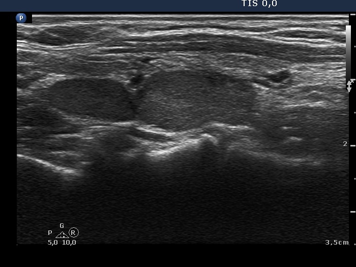
Right lobe, longitudinal scan. The thyroid presents fibrosis. Compared with the previous examination, the lower hypoechogenic mass increased in size.