
|
|
Ethanol sclerotherapy: non-toxic solid nodules - Case 3
|
|


First session of therapy (first row of images)
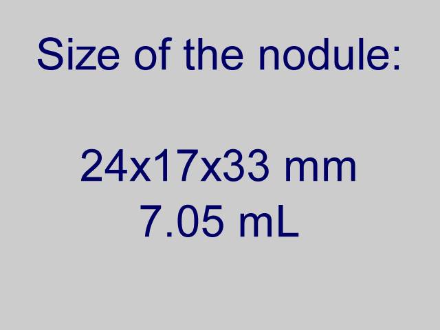
Clinical presentation. A 65-year-old colleague was regularly checked for more than a decade because of a nodule in the left lobe. At first visit the size of the nodule was 15x11x22 mm. Except for cosmetics the nodule did not cause complaint.
Palpation: a firm nodule in the left lobe.
Functional state: euthyroidism with TSH 1.85 mIU/L.
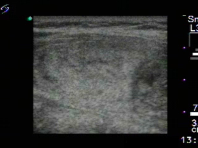
Ultrasonography: The thyroid was echonormal. A large nodular area replaced the normal parenchyma in the left lobe. The lesion was composed of a larger echonormal nodule presenting halo sign and a smaller hypoechogenic one.
Aspiration cytology of the hypoechogenic nodule resulted in benign cystic-colloid goiter.
The colleague wished to avid surgery and decided to undergo sclerotherapy instead. 5 sessions of sclerotherapy were performed. On the first occasion, we injected 3 mL ethanol.
Third session of therapy (second row of images)
At this time we gave 1.5 mL ethanol.
Fifth session of therapy (fourth row of images)
At this time we administered 3 mL ethanol. This was the last session, the patient get 11 mL ethanol in the aggregate.
Seven years after the therapy (fourth row of images)
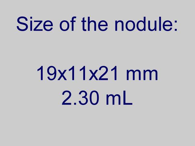
Clinical presentation: the patient underwent yearly ultrasound and TSH checking. The volume of the nodule remained less than 30% of the original size in all visits. She had no complaints.
Palpation: a nodule in the right lobe.
Functional state: euthyroidism with TSH 1.07 mIU/L.
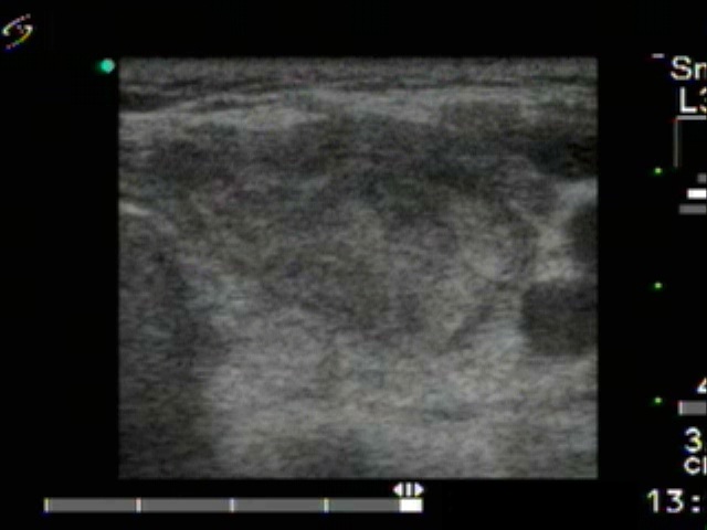
Ultrasonography: The thyroid was echonormal. The nodular area became hypoechogenic and significantly smaller.