
|
|
Ethanol sclerotherapy: toxic nodules - Case 1
|
|


Clinical presentation: A 57-year-old man with an autonomously functioning nodule known for 10 years was referred for an evaluation. Previous hormonal examinations indicated euthyroidism. 4 months before the present visit, he got iodine containing contrast material. 6 weeks later hyperthyroidism had developed (TSH 0.001 mIU/L, FT4 51.1 pM/L). Despite large dose of thyrostatic therapy, the hyperthyroidism did not improve over 1 0 weeks.
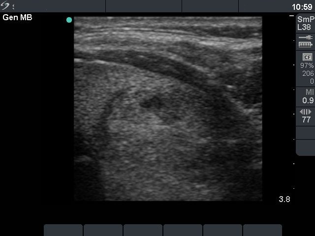
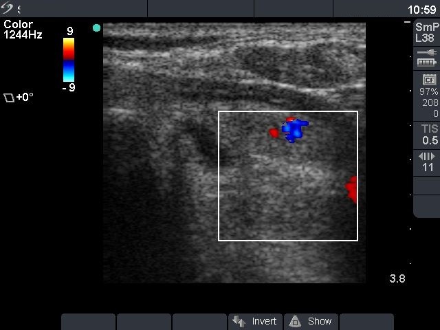
First row: ultrasonographic picture before sclerotherapy. There was an echonormal nodule in the left lobe with the dimensions of 30x27x31 mm (width x depth x length). 4 sessions of ethanol sclerotherapy were administered.
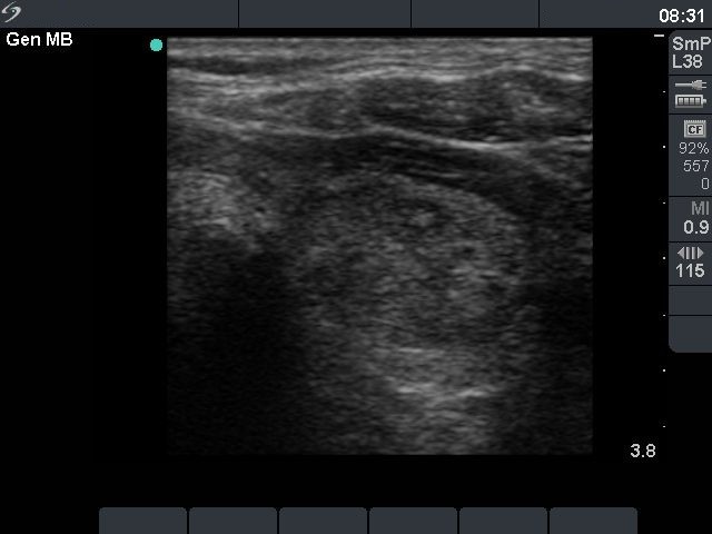
Second row: 6 weeks after the last session. The FT4 levels decreased to 39.1 pM/L on daily 30 mg methimazole. The dimensions of the nodule were 21x14x27 mm (width x depth x length). 6 weeks later hypothyroidism has developed. The methimazole-therapy was discontinued.
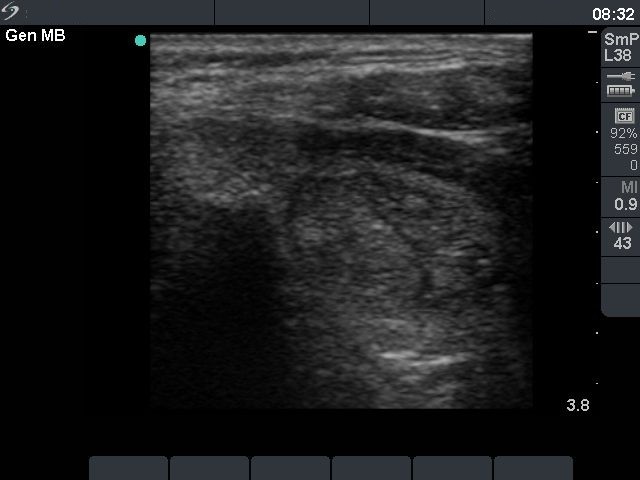
Third row: 10 months after sclerotherapy. The patient was clinically and biochemically euthyroid (TSH 1.09 mIU/L, FT4 15.2 pM/L). The dimensions of the nodule were 19x14x24 mm (width x depth x length).