
|
|
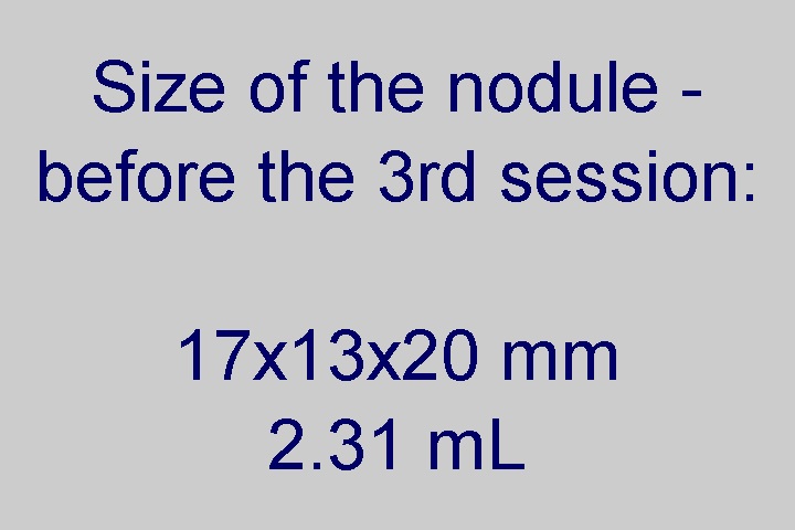
Ethanol sclerotherapy: other examples - Case 4: therapy of a pregnant patient
|
|


Three years before the therapy (first row of images):
Clinical presentation: A 34-year-old man was referred for an evaluation of a recurrent thyroid nodule. She underwent subtotal lobectomy of the right lobe 7 years ago. Histopathology disclosed follicular adenoma. She noticed a lump several weeks before the examination.
Palpation: a large elastic nodule in the left lobe.
Functional state: euthyroidism with TSH 2.61 mIU/L.
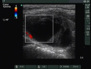
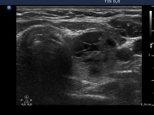
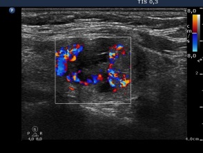
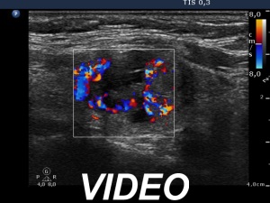
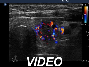
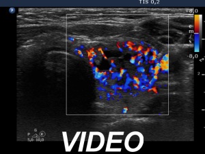
Ultrasonography: The thyroid was echonormal. There was a dominantly cystic nodule in the left lobe. The lesion displayed signs of perinodular and intranodular blood flow.
Aspiration cytology was performed and we gained 8 mL brown cystic fluid. The cytological report was benign cystic lesion.
First session (second row of images):
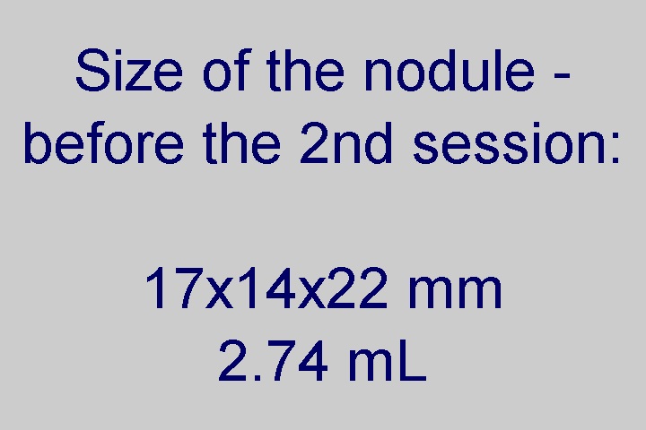
Summary of the previous years: Tthe cyst has refilled within 2 weeks. At this time 9 mL brown fluid was aspirated. The patient was regularly checked. The size of the nodule remained much smaller in the previous 3 years, the dimensions of the lesion was 16x16x26 mm at the last follow-up three month ago.
Clinical presentation: The patient became pregnant, she was at the 13th gestational week. She noticed a significant increase in the nodule size during the pregnancy which caused discomfort recumbent position. I offered ethanol sclerotherapy.
I aspirated 0.3 mL brown fluid thereafter injected 1.5 mL ethanol into the nodule. At the end of the session the patient had a burning pain lasting for a minute.
Second session (third row of images):
Clinical presentation: The patient became aware of mild neck discomfort lasting for 2 days.
I administered 1.5 mL ethanol. The treatment caused a burning pain lasting for 30 seconds.
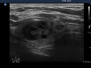
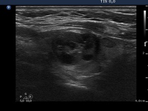
Third session (fourth row of images):
Clinical presentation: The patient had no complaints.
I injected 1.5 mL ethanol. The patient had no complaints.
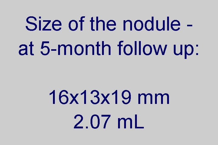
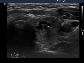
Five months after the last session (fifth row of images):
Clinical presentation: The patient had no complaints.
Palpation: no abnormality.
Functional state: euthyroidism with TSH 1.88 mIU/L.
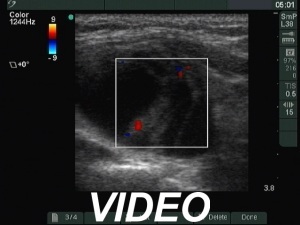
Ultrasonography. The nodule became much smaller. The increased intranodular vascularization remained unchanged.
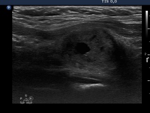
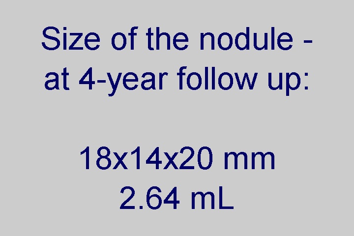
Four years after the last session (sixth row of images):
Clinical presentation, palpation, functional state (TSH 1.09 mIU/L) were unchanged.
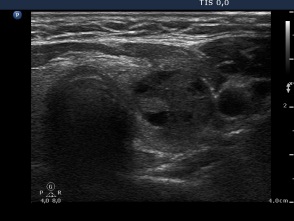
Ultrasonography revealed a minimal-moderate increase in nodule size. Compared with the previous examination the solid part has increased. The vascularization remained significantly increased.