
|
|
Aberrant thyroid - case 1150
|
|


Clinical data: A 38-year-old woman was referred for evaluation of a nodular goiter. The patient noticed a lump in the middle part of the neck.
Palpation: a not firm mass in the middle part of the neck above the level of the thyroid.
Functional state: euthyroidism with TSH 0.84 mIU/L.
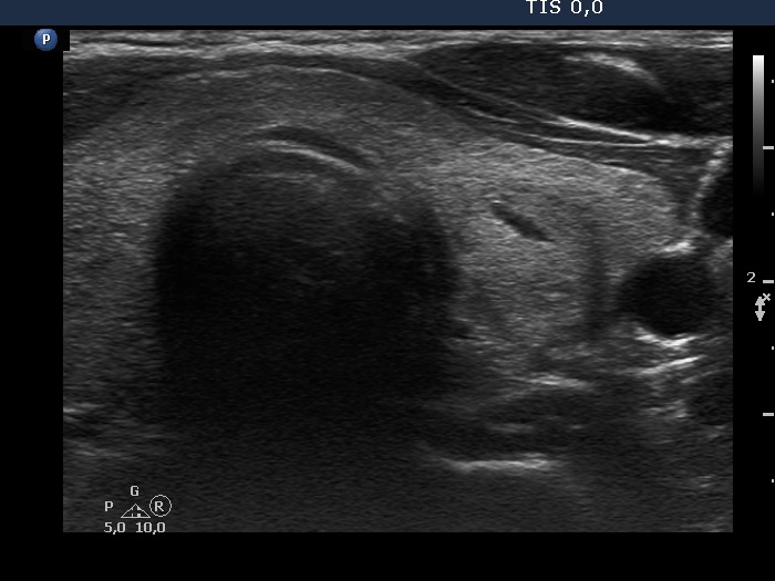
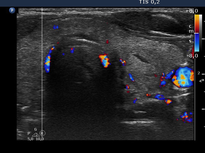
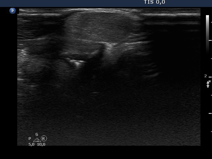
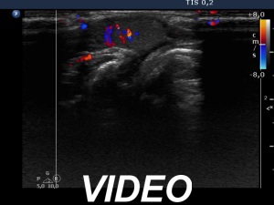
Ultrasonography. The thyroid was echonormal. Both lobes had an echonormal nodule which showed halo sign. The left had perindoular blood flow while the left one presented taller-than-wide shape. There was a moderately hypoechoic mass in the middle part of the neck. This lesion had intralesional vascularity.
Cytology was performed from all three lesions. Cytology resulted in benign follicular proliferation, colloid goiter and colloid goiter, right lobe, left lobe, aberrant tissue.
Suggestion ultrasound in two years.
Comment. The cytology would not be indicated on TIRADS in the event of the right nodule because the largest diameter of the lesion was 19 mm. Nevertheless, the ultrasound pattern suggested follicular tumor and the FNA was also consistent with this. The published data suggest a 15-25% risk of malignancy in the event of a cytologically suspected follicular tumor. This ratio is much lower in Hungary, and specifically in my practice it is around 3%. In this patient, normofollicles predominated the smear, which decreases the malignant version of a follicular tumor.