
|
|
Aberrant thyroid - case 952
|
|


Clinical data: A 41-year-old woman was referred for evaluation of a repeatedly elevated TSH which ranged between 3.2 and 5.8 mIU/L in the last three years. She had no complaints and did not receive replacement therapy because she did not wish to be pregnant. Recently, she changed her mind and wished to be pregnant.
Palpation before the ultrasound: no abnormality.
Laboratory tests: TSH 4.91 mIU/L, FT4 15.8 pM/L, aTPO 1.0 U/mL.
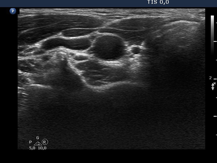
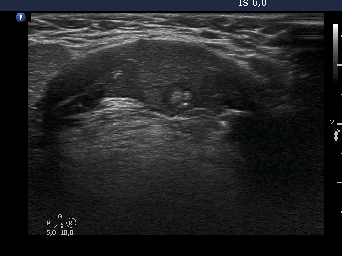
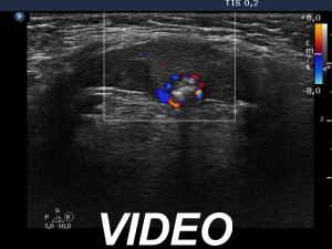
Ultrasonography. There was no thyroid tissue in the usual location. Considering the only minimal degree of hypothyroidism, we searched for a possible thyroid tissue in the neck. We have found a minimally hypoechoic discrete area within the submental muscle. The dimensions of this area were 13x11x16 mm, width, depth, length, respectively. There was a hypoechoic discrete lesion in this area. The discrete lesion had amorphic echogenic fragments and punctate echogenic foci, too.
After the ultrasound examination, we repeatedly palpated the neck of the patient. A firm lesion was found in the submental area.
On aspiration cytology of both the minimally hypoechoic area and the discrete lesion within, we have found typical epithelial cells in regular, small sheets. There was neither colloid nor lymphoid cell population on the smears.
Wash-out thyroglobulin exceeded 478 microgram/L.
Suggestion: daily 50 microgram levothyroxine, TSH in 3 months, ultrasound in 2 years.
Scintigraphy was indicated which showed radioiodine uptake only according to the submental mass.
Comment.
-
The lesion is clearly an ectopic thyroid.
-
The discrete lesion within has a suspicious appearance and although cytology was benign, we intend to repeat FNA later in the course.