
|
|
The issue of large goiters - case 757
|
|


Clinical data: A 37-year-old woman was referred for evaluation of a nodular goiter which was discovered by the patient herself.
Palpation: The right lobe was enlarged and a not firm nodule was palpated within.
Laboratory tests: TSH 0.26 mIU/L, FT4 15.8 pM/L.
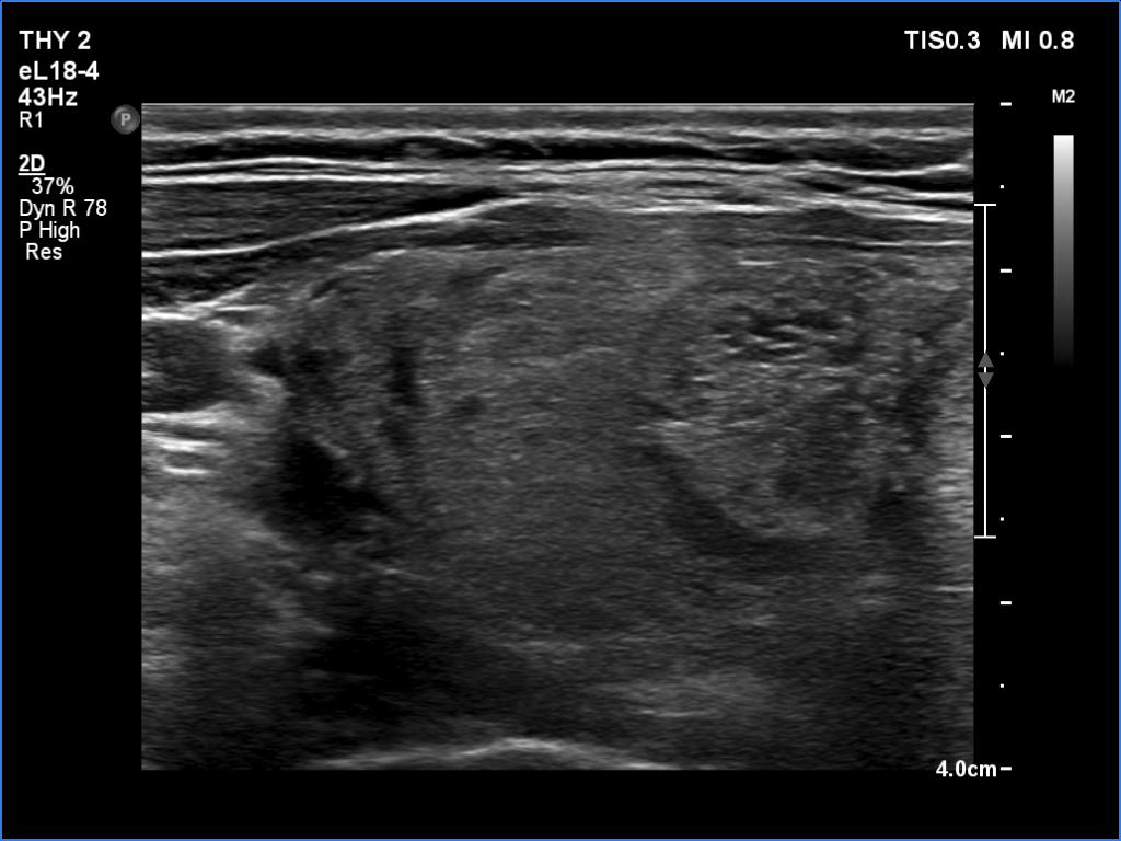
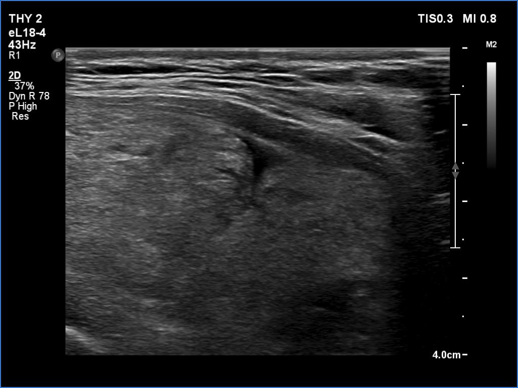
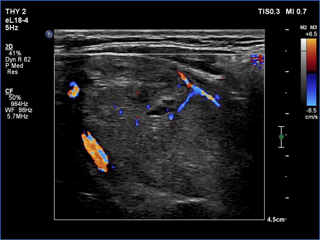
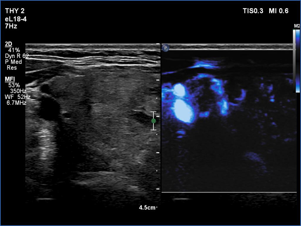
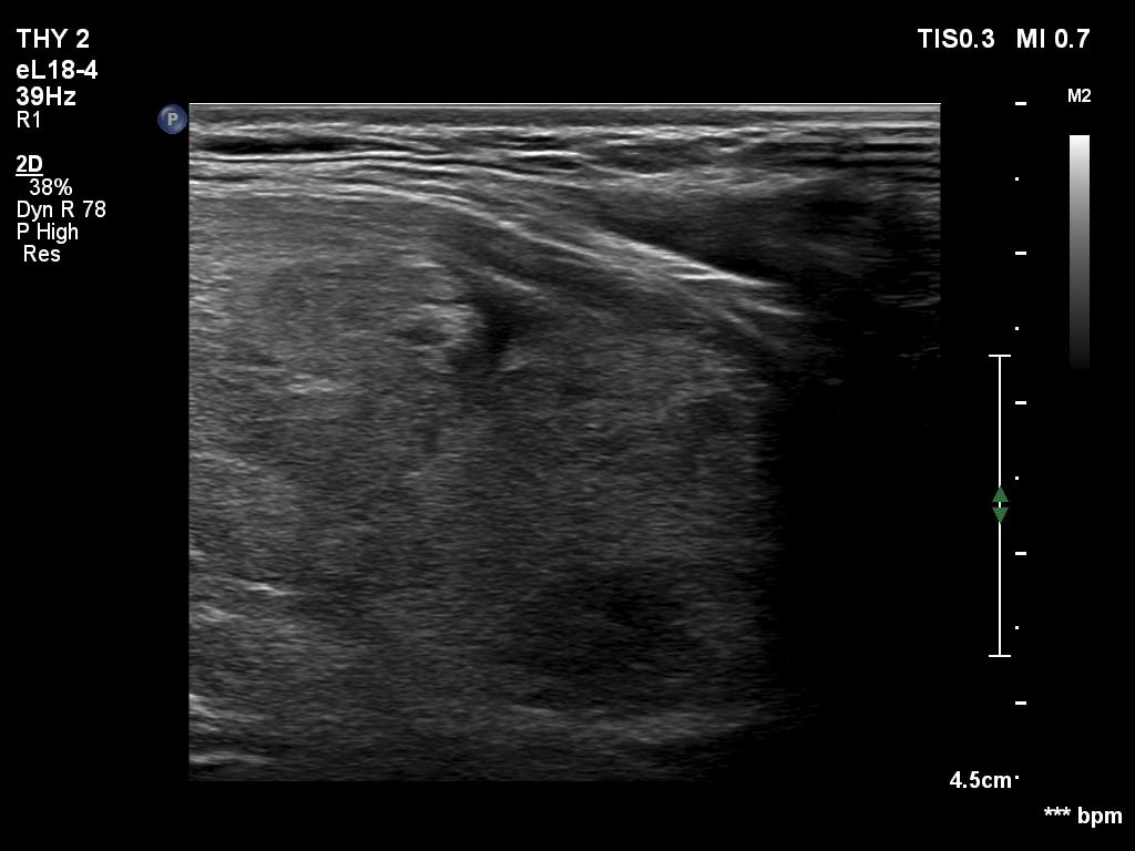
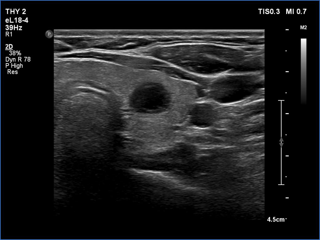
Ultrasonography. The thyroid was echonormal. The right lobe was composed of multiple nodules with different echogenicities. The lower pole of the right lobe could not be visualized in a supine position with the neck held back neither before nor while swallowed. However, at the latter occasion, technical reasons hindered the judgement of the lower pole.
Cytology resulted in benign follicular proliferation.
Right lobectomy was performed. Histopathology disclosed benign hyperplastic nodules.
Comment. If the lower pole of an enlarged lobe becomes clearly visible during swallowing, then it can be safely said that the thyroid gland can be removed by neck excision. If we have any doubt, a neck and upper mediastinal CT scan is mandatory.