
|
|
The issue of large goiters - case 902
|
|


Clinical data: A 40-year-old man was referred for evaluation of a nodular goiter which was discovered on ultrasound screening. an evaluation after his twin-brother was diagnosed with papillary carcinoma. He had no complaints.
Palpation: The left lobe had a not firm nodule.
Laboratory tests: TSH 3.01 mIU/L, FT4 13.1 pM/L.
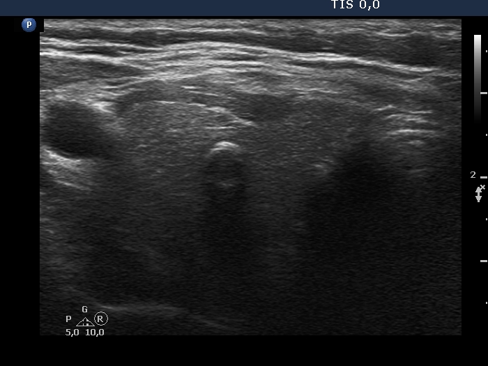
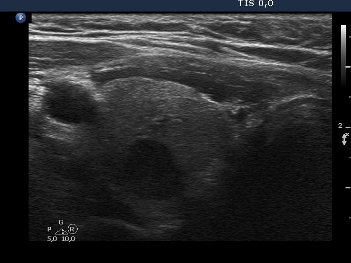
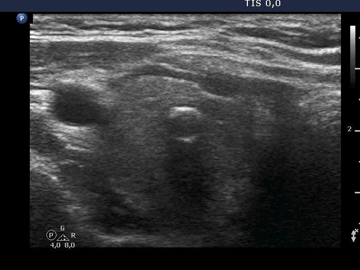
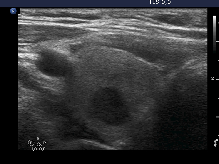
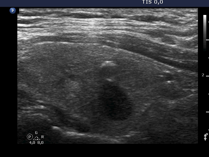
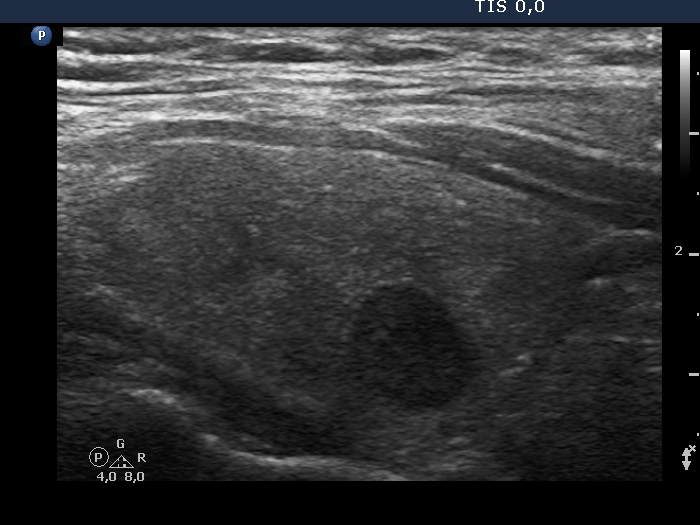
Ultrasonography. The right lobe was minimally while the left one was deeply hypoechoic. There were multiple lesions in the right lobe, including a small one having macrocalcification and a deeply hypoechoic nodule. There was a nodular mass in the lower half of the left lobe. The mass was composed of three discrete echonormal lesions.
Additional laboratory test: aTPO 206 U/mL.
Cytology was performed form the hypoechoic lesion in the right lobe and from the echonormal nodular mass in the left lobe. The FNA of the former resulted in lymphocytic thyroiditis while the cytology of the latter did in benign colloid goiter.
Comments.
-
It is worth comparing the influence of the setting on the image quality and on the presentation of ventral and dorsal structures. (See images and video of the right lobe.)
-
It is unusual for the two lobes to have different echo patterns.