
Case 116 (ultrasonographic picture 4)
|
|

|
|
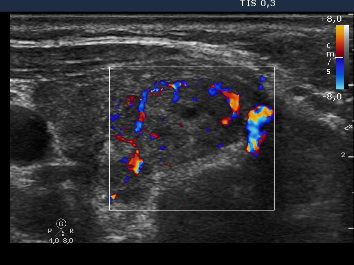
Right lobe, transverse view, color Doppler mode. Type 2 vascular pattern, i.e. the lesion displays perinodular blood flow.
|
|
|
|
Right lobe, transverse view, color Doppler mode. Type 2 vascular pattern, i.e. the lesion displays perinodular blood flow.