
The operated thyroid - case 49 (ultrasonographic picture 5)
|
|

|
|
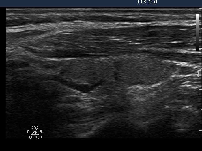
Left lobe, longitudinal scan. The lobe seems to be consisted of two or three parts. However, the echogenicity of these areas is identical, and only the deeply hypoechoic vascular and the moderately hypoechoic fibrous structures make the appearance nodular.