
|
|
Thyroid cancers - case 1060
|
|


Clinical presentation: A 79-year-old man was referred for aaspiration cytology. He noticed a rapidly increasing nodule causing dysphagia and hoarseness. The goiter evolved within a several weeks.
Functional state: euthyroidism with TSH-level 3.73 mIU/L, FT4 16.0 pM/L.
Ultrasonography. The right lobe was intact, while the left lobe was replaced by a very large hypoechogenic mass. A large vessel was found within the nodule.
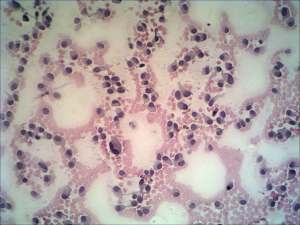
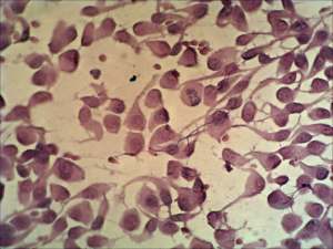
Cytological picture. There was no colloid in the background. The smear was extremely cellular. The cells formed neither follicles nor papillary fronds, they were arranged in loose syncytial structures and predominantly dissociated. The atypical cells were polygonal with eccentric nuclei. There were numerous triangular forms specific for medullary cancer and several highly atypical, pleomorphic tumor cells.
Cytological diagnosis: medullary cancer.
Blood test for calcitonin: serum-level of calcitonin exceeded 585 pM/L (normal value: 0-3.36).
Histopathology disclosed medullary cancer.