
Study on 100 consecutive patients with thyroid nodule - case 001 (cytologic picture 1)
|
|

|
 |
|
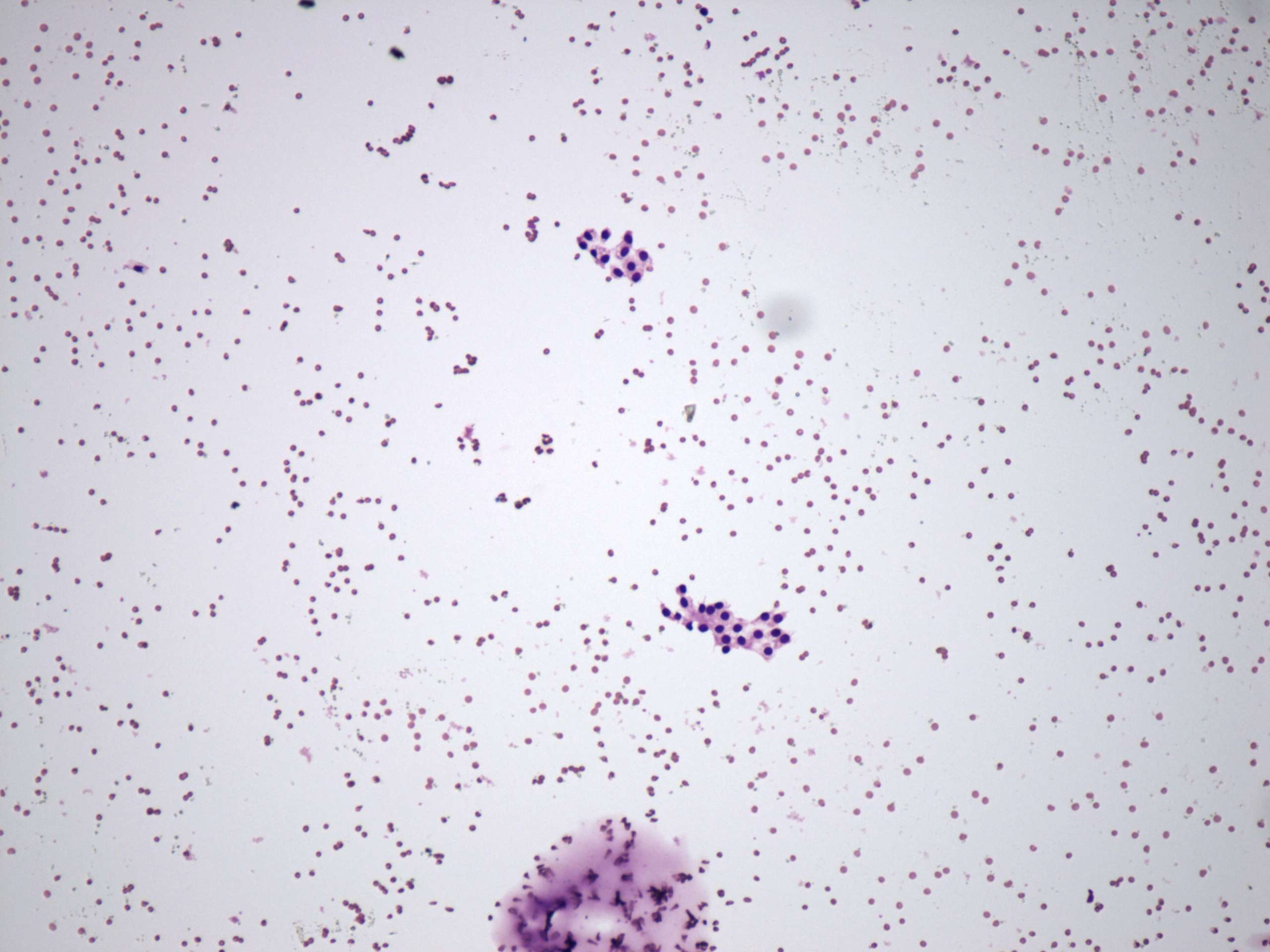
Wright-Giemsa staining, 100x. There is a colloid spot in the lower part of the image. Two groups of microfollicles can be found.