
|
|
Study on 100 consecutive patients with thyroid nodule - case 002
|
|


Clinical presentation: A 63-year-old woman was referred for aspiration cytology. A multinodular goiter was revealed on ultrasound screening.
Palpation: a moderately firm nodule in the isthmus.
Functional state: euthyroidism (TSH 1.51 mIU/L).
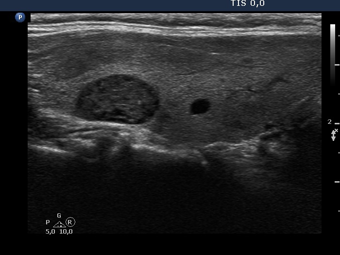
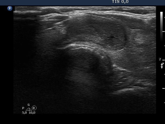
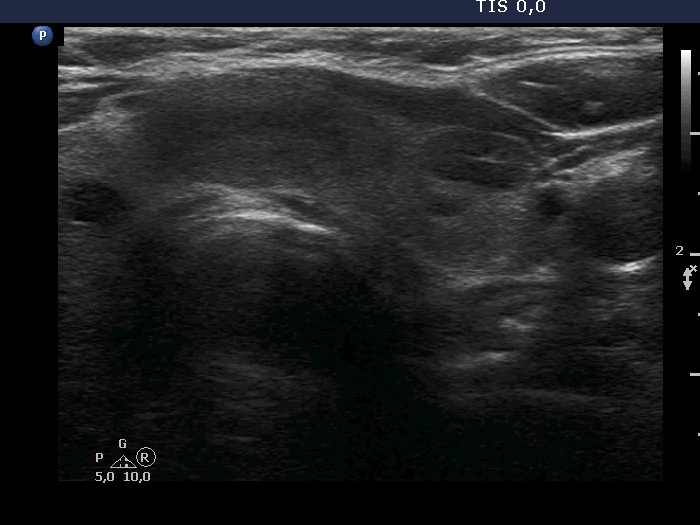
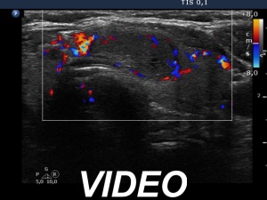
Ultrasonography. The thyroid was echonormal and had several nodules. There was a hypoechoic lesion in the right lobe which presented tiny cystic areas. Another moderately hypoechoic nodule in the isthmus showed lobulated margins, had halo and perinodular blood flow. Video proves that this nodule had punctate echogenic foci, too. The left lobe contained multiple lesions including a hypoechoic one which presented a microcalcification.
FNA was performed from the nodule in the isthmus and from the hypoechoic lesion in the left lobe that had microcalcification. Cytology resulted in benign lesion in both nodules.
Suggestion: ultrasound in two years.