
Study on 100 consecutive patients with thyroid nodule - case 011 (ultrasonographic picture 1)

|

|
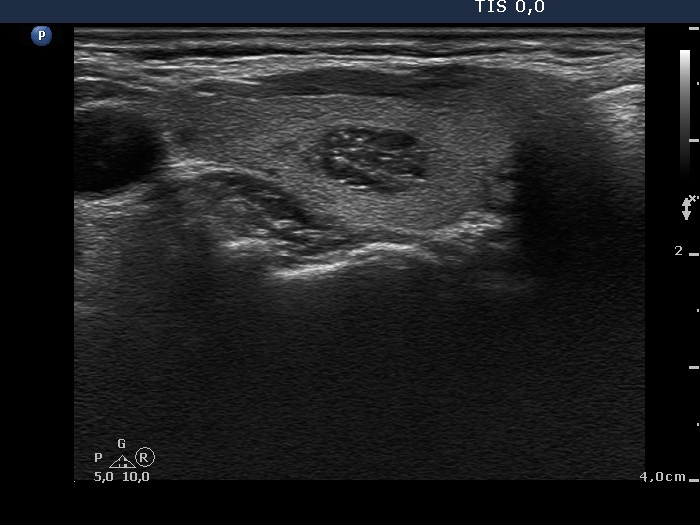
Right lobe, transverse scan. There is a hypoechogenic nodule in the central part of the lobe. The lesion shows intranodular hyperechogenic figures, the hyperechogenic granules and lines correspond to fibrosis or to back wall figures.


