
Study on 100 consecutive patients with thyroid nodule - case 013 (ultrasonographic picture 7)

|

|

|
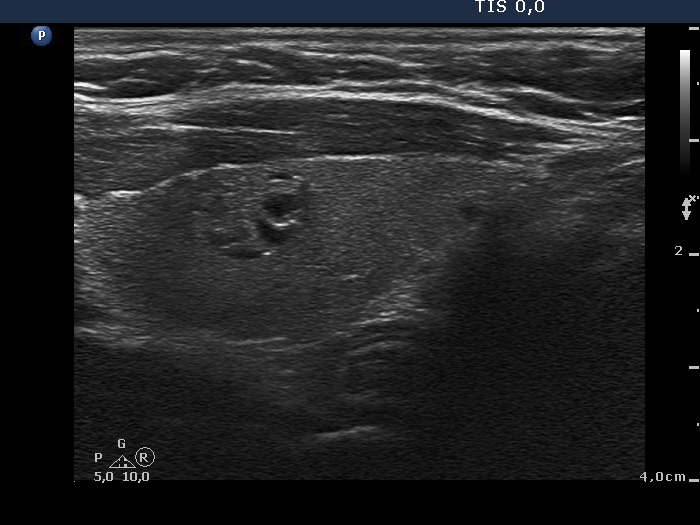
Left lobe, longitudinal scan. The bright hyperechogenic intranodular figures are not microcalcifications but signs of fibrotic changes or back wall figures, because they are in part linear and are related to ventral cystic areas.


