
|
|
Study on 100 consecutive patients with thyroid nodule - case 016
|
|


Clinical presentation: A 65-year-old woman was referred for evaluation of a thyroid nodule discovered on PET-CT. She has been operated on colon carcinoma for 6 months. The PET-positive lesion was found in the right thyroid.
Palpation: The surface of the right lobe was uneven. No nodule could be palpated.
Functional state: euthyroidism (TSH 2.51 mIU/L).
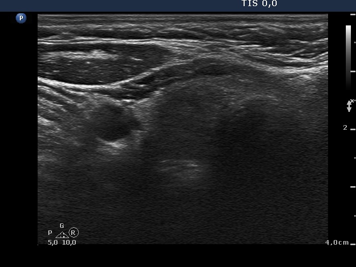
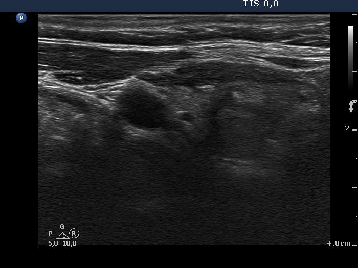
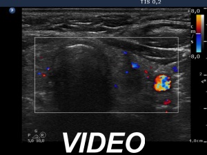
Ultrasonography. The thyroid was echonormal-minimally hypoechogenic. There were multiple discrete lesions in the right lobe. None of them presented suspicious echo pattern. There was a two-chambered cyst in the left thyroid.
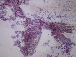
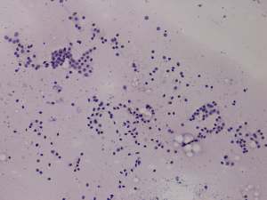
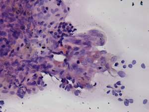
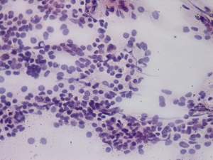
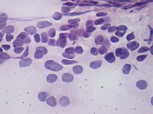
Cytology was performed from two lesions located in the right lobe. Cytological diagnosis: benign colloid goiter.
We performed anti-TPO test which yielded 2 U/mL.
Comment. The echo pattern resembles that of a Hashimoto's thyroiditis both because of the basic hypoechogenicity and the pattern of the lesions in the right lobe. Nevertheless, neither the cytology nor the anti-TPO level was positive for an autoimmune process.