
|
|
Study on 100 consecutive patients with thyroid nodule - case 041
|
|


Clinical presentation: A 35-year-old woman came to a follow-up. First, we examined her 4 years ago. At this time two nodules were found in the left lobe. The dimensions of the lower lesion were 23x20x25 mm. Cytology of this nodule resulted in follicular tumor without atypia. The patient did not want to be operated. Therefore, a yearly ultrasound check was suggested.
Palpation: The left lobe was nodular on palpation with a not firm lesion.
Functional state: euthyroidism (TSH 1.86 mIU/L).
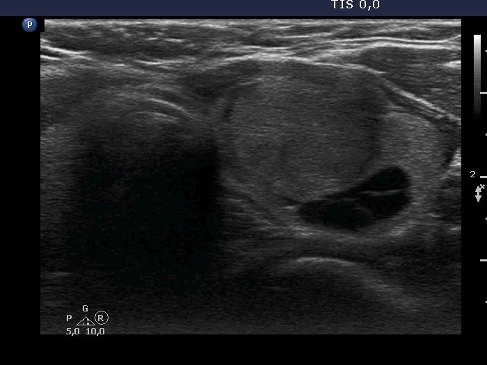
Ultrasonography. The thyroid was echonormal. There were two nodules in the left lobe. The upper was a dominantly cystic lesion while the lower was a dominantly solid, heterogeneous nodule with a dominant hypoechoic part. The dimensions of the nodule were 23x20x24 mm. The nodule had halo and perinodular vascularity.
Suggestion: The decision on surgery can still be postponed with annual review.
Comment.
-
The tumor did not present oxyphilic changes. The oxyphilic cells presented in the cytological images might arise partly from the extranodular part of the lobe which presented Hashimoto's thyroiditis.
-
The nodules present all three types of irregular margins, the borders are partly blurred and have lobulations and spiculations, as well.