
|
|
Study on 100 consecutive patients with thyroid nodule - case 046
|
|


Clinical presentation: A 24-year-old woman was referred for evaluation of a hypothyroidism discovered on screening. She had no complaints.
Palpation: Both lobes were firm. The isthmic part of the right lobe was hard. It was doubtful whether there is a nodule or not.
Hormonal examination: subclinical hypothyroidism with TSH-level 6.31 mIU/L, FT4 15.8 pM/L, aTPO 317 U/mL.
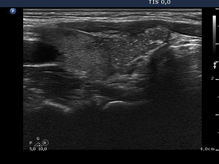
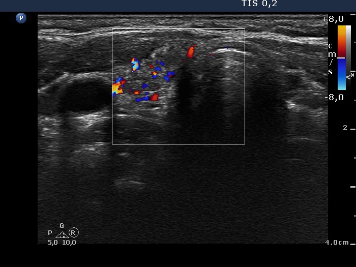
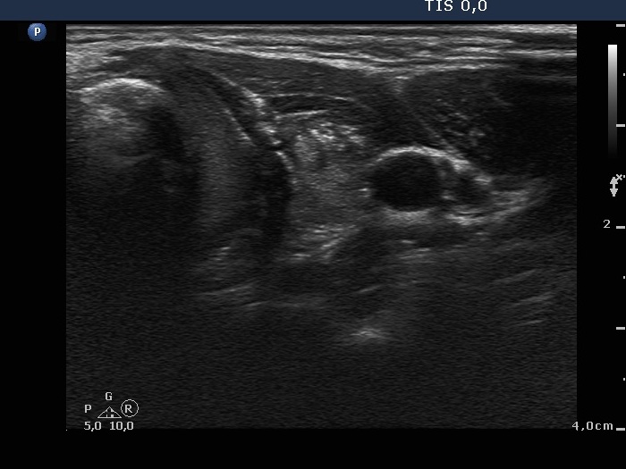
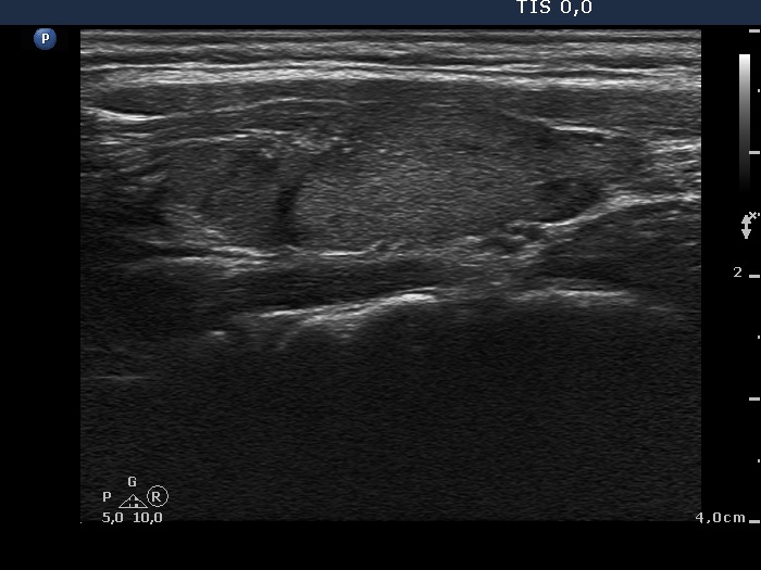
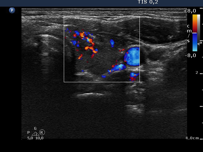
Ultrasonography. The thyroid was minimally hypoechogenic and contained several more hypoechogenic areas. There were two hypoechogenic nodules, one in the isthmic part of the right lobe and another one in the ventral part of the left lobe. Both lesions presented the so-called starry sky phenomenon, i.e. they contained numerous microcalcifications. The nodules had irregular, partly lobulated, partly invasive margins. The lesions displayed increased intranodular blood flow, in the event of the left nodule a chaotic, irregular vascularization was found.
Cytology was performed. Two samples were gained from the lesion in the right lobe. The patient refused to aspirate the nodule in the left lobe.
Considering the ultrasound presentation, we told the patient that irrespectively from the result of the cytology surgery cannot be avoided.
One hour later we made the microscopic analysis. We found only blood cells on the smears.
We offered the patient a repeat aspiration. Taking our opinion into account the patient decided not to repeat the aspiration.
Final diagnosis: subclinical hypothyroidism caused by Hashimoto's thyroiditis. Suspicion of multifocal papillary carcinoma with a more than 95% risk of malignancy.
Surgery was performed. Intraoperative frozen section disclosed papillary carcinoma. Histopathology: multifocal papillary carcinoma in both lobes. Lymphocytic thyroiditis.
Comments.
-
Such ultrasound presentation (starry sky phenomenon, chaotic type vascular pattern, irregular borders in a hypoechogenic nodule) is almost itself diagnostic for a papillary carcinoma. The risk being such lesion a papillary carcinoma is more than 95%.
-
This case illustrates the difficulty to gain adequate material from a vascularized tumor.
-
In the everyday practice it is not always possible to follow the recommendations, and I think that in such situations to resign from a repeat FNAC was a correct decision.
-
And first of all, this case illustrates the systematic need for a new way of thinking: in contrast with the current protocol, the role of ultrasound should not be only the selection of nodules for cytology but must have play a role even in the final diagnosis.
- The lesions presented partly lobulated margins. But the invasive borders were more important in this case. This type of border is characterized by a central core presenting microcalcifications and isolated microcalcification relatively far from the core.