
|
|
Study on 100 consecutive patients with thyroid nodule - case 051
|
|


Clinical presentation: A 40-year-old woman came to a follow up. We met her for the first time 8 years ago because of a nodular goiter that grew slowly over the years. Cytology was benign, no hormonal abnormalities have been observed so far.
Palpation: a not firm nodule in the left lobe.
Functional state: subclinical hyperthyroidism (TSH 0.04 mIU/L, FT4 15.1 pM/L, FT3 6.47 pM/L).
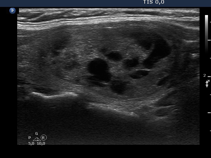
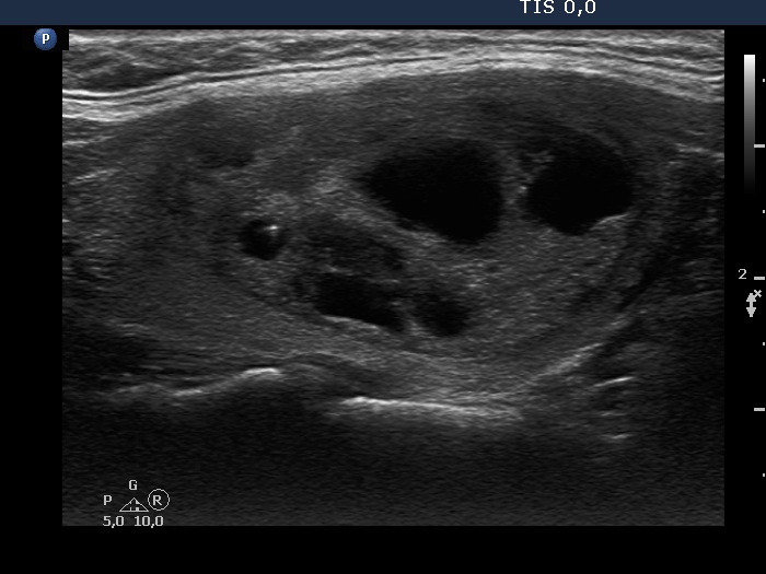
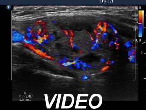
Ultrasonography. The thyroid was echonormal. There was a dominantly solid nodule in the right lobe. The lesion has halo and showed both perinodular and increased intranodular blood flow.
Scintigraphy disclosed an autonomously functioning adenoma.
Radioiodine therapy was advised.
Comment. This case illustrates all typical ultrasound features of an autonomously functioning adenoma causing hyperthyroidism. First, there is a relatively large solitary. Second, it shows the characteristic ultrasound signs of a follicular tumor (halo sign and perinodular blood flow). There are two more, important signs in the hyperthyroid state: the intranodular blood flow is usually increased while the other lobe becomes decreased in size because of the low TSH level.