
Study on 100 consecutive patients with thyroid nodule - case 067 (ultrasonographic picture 1)

|
|
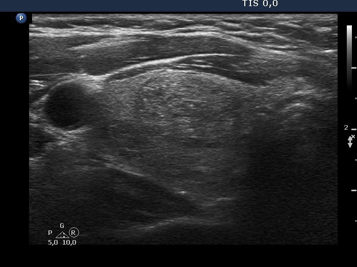
Right lobe, transverse view. There is a minimally hypoechogenic, inhomogeneous nodule in the ventral half of the lobe. The lesion has both echogenic lines and granules. These figures are presentations of connective tissue.