
|
|
Discrete lesion or nodule in Hashimoto's thyroiditis - case 21 (1496)
|
|

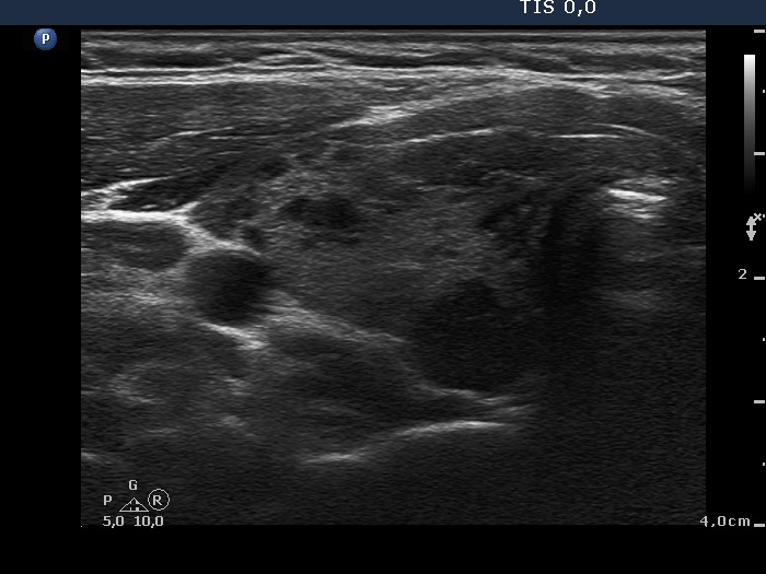


Clinical presentation: A 22-year-old woman was referred for evaluation of a thyroid nodule detected on evaluation of amenorrhoea. One of the lesions located in the dorsal part of the right lobe was described as a "highly suspicious", microlobulated, TIRADS 5 category nodule. The patient was examined because of suspected thyroid enlargement at the age of 14 when I found numerous hypoechogenic areas within an echonormal background.
Palpation: Both lobes were a bit firm. No nodules could be palpated.
Laboratory tests: TSH 2.07 mIU/L, aTPO 394 U/mL.
Ultrasound. The thyroid was echonormal and presented numerous hypoechogenic areas. Neither the shape nor the borders of these hypoechogenic fields corresponded to true nodules. The lesion in question did not differ from other hypoechogenic areas.
Aspiration cytology: Hashimoto's thyroiditis.
Comments.
-
The lesions, including the aspirated one, are unlikely being nodules in pathological sense.
-
The pattern, including the lobulated/spiculated margins of the discrete lesions, is specific for Hashimoto's thyroiditis.