
|
|
100 consecutive cases of papillary cancer - case 028
|
|


Clinical presentation: A 59-year-old woman was referred for aspiration cytology. She has been treated for breast cancer for two years. On PET CT scan a nodule in the left thyroid was detected.
Palpation: a hard nodule in the ventromedial part of the left lobe.
Functional state: euthyroidism (TSH 0.99 mIU/L).
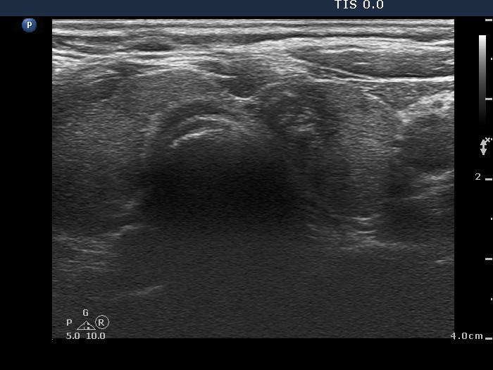
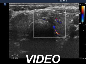
Ultrasonography. The thyroid was echonormal. There was a hypoechogenic nodule in the ventromedial part of the left lobe. The lesion presented hyperechogenic figures which corresponded to proliferation of connective tissue. The simultaneous presence of microcalcifications was equivocal.
Cytology resulted in papillary carcinoma.
Histopathology disclosed follicular variant of papillary carcinoma. The maximal diameter of the tumor was 8 mm.